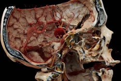
Researchers at the University of Zurich in Switzerland say they have produced the first cinematically rendered image from a patient based on a standard clinical F-18 FDG-PET/MRI scan.
The 3D image is from a patient with an oropharyngeal carcinoma in the right-sided soft palate and palatine tonsil, noted first author and nuclear medicine physician Martin W. Huellner, MD, and colleagues.
"Historically, [cinematic rendering] has primarily centered on CT images, with sporadic instances noted in PET/CT; however, its use in PET/MR remains unreported to date," the group wrote. The article was published on July 1 in the European Journal of Nuclear Medicine and Molecular Imaging and featured as the journal's Image of the Month.
 Signa PET/MR scanner (GE HealthCare, Waukesha, WI) MR image: T1-weighted LAVA-flex, repetition time, 4.65 ms; echo time, 1.82 ms; flip angle, 15°; slice thickness,1.3 mm; spacing, 2.6 mm; field of view; 100 mm; total acquisition time; 1:31 minutes. PET image: 245 MBq of F-18 FDG, bed time, 1:30 minutes; reconstructed using block-sequential regularized expectation maximization. Image courtesy of Huellner et al. Licensed under CC BY 4.0.Image courtesy of the European Journal of Nuclear Medicine and Molecular Imaging.
Developed as a fusion of radiology and cinematography, cinematic rendering transcends traditional rendering methods and provides lifelike representations that bridge the gap between medical images and reality, the authors wrote. It is a postprocessing technique based on the segmentation of standard images into photorealistic 3D representations, facilitating spatial orientation.
The image was retrospectively reconstructed from a standard F-18 FDG-PET/MRI clinical image dataset acquired from the patient within less than two minutes of scan time using the software Cinematic Anatomy (Siemens Healthineers, Erlangen, Germany), they noted.
"Such photorealistic images could potentially aid radiologists in reporting and surgeons in preoperative planning by offering crucial anatomical landmarks, enhancing visualization of subsurface structures, and complementing endoscopic images," the group concluded.
Huellner's co-authors included Grégoire Morand, MD, of the department of otolaryngology-head and neck surgery; Bernd Stadlinger, MD, of the Clinic of Cranio-Maxillofacial and Oral Surgery; and Klaus Engel, PhD, of Siemens Healthineers.
The full article is available here.


![Current PET/MRI imaging of healthy and damaged mouse kidneys using the dual contrast agent F-18 [Gd (FL1)]. The imaging provides consistent signals from both PET and MRI. In healthy kidneys, the center of the kidneys appears dark in the MRI image and very bright in the PET image, indicating normal excretion of the contrast agent. The onset of impaired function in one kidney is indicated by the accumulation of the contrast agent and its slow excretion. Precise mapping of MRI and PET data ensures detailed morphological and quantitative analysis of kidney impairment. Image and caption courtesy of IOCB Prague.](https://img.auntminnie.com/files/base/smg/all/image/2024/08/PET_MRI_agent.66b25358df3b3.png?auto=format%2Ccompress&fit=crop&h=167&q=70&w=250)