F-18 fluorocholine (FCH) PET/CT could be a first-line technique over technetium-99m sestamibi (Tc-99m MIBI) SPECT/CT for imaging parathyroid tumors prior to surgery, according to a study published June 20 in JAMA Otolaryngology-Head & Neck Surgery.
The recommendation is from a phase III trial in France that compared the approaches to determine optimal care in patients with primary hyperparathyroidism (PHPT), noted lead author Elske Quak, MD, of the Centre François Baclesse in Caen, and colleagues.
“These findings suggest that first-line FCH-PET/CT can replace MIBI-SPECT/CT for imaging-guided surgery in patients with PHPT,” the group wrote.
Medical imaging is used to localize parathyroid adenomas in patients with PHPT, a condition often diagnosed based on high calcium levels. The imaging allows surgeons to perform minimally invasive parathyroidectomies instead of more invasive procedures, the authors explained.
MIBI-SPECT/CT is considered the standard technique for guiding these surgeries, yet several phase II clinical trials suggest advantages of a newer approach using FCH-PET, such as superior spatial resolution for detecting small adenomas and lower radiation exposure to patients, they noted.
To establish which of the two is the optimal first-line approach, the group enrolled 57 patients (63 years old on average; 15 men and 42 women) in a multicenter randomized diagnostic intervention trial.
Between November 2019 and May 2022, patients received first-line FCH-PET/CT (n = 29) or MIBI-SPECT/CT (n = 28) prior to surgery. After one month, the researchers compared how successful the surgeries were among the two groups based on serum calcium levels corresponding to the upper limit of normal.
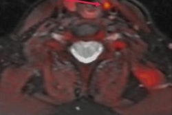
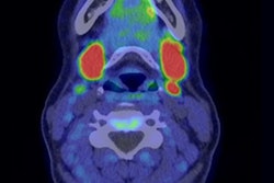
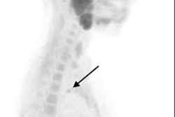
 (A) FCH-PET maximum-intensity projection; (B) FCH-PET/CT fusion transverse slice; and (C) low-dose CT transverse slice, showing high FCH uptake in an 11-mm left inferior parathyroid adenoma (arrowheads). The patient underwent minimally invasive parathyroidectomy, leading to normocalcemia during follow-up. No complications occurred.Image courtesy of JAMA Otolaryngology-Head & Neck Surgery
The primary endpoint was assessable in 27 patients in the PET/CT group and 25 patients in the SPECT/CT group. The researchers found normocalcemia one month after FCH-PET/CT-guided surgery in 23 patients (85%) and in 14 patients (56%) one month after MIBI-SPECT/CT, according to the results.
The sensitivity was 82% for FCH-PET/CT and 63% for MIBI-SPECT/CT, the authors reported.
“FCH-PET/CT leads more patients with PHPT to correct imaging-guided MIP and normocalcemia than MIBI-SPECT/CT thanks to its superior sensitivity,” the group wrote.
FCH was first developed in the early 2000s as a PET radiotracer for detecting prostate cancer and staging patients. Additionally, the tracer was found useful in 2014 in patients with PHPT. This study further supports its use in these patients, the team wrote.
“We expect these findings to contribute to the marketing authorization of this existing radiopharmaceutical for a new clinical indication,” it concluded.
The full study is available here.