Europe
Clinical News
Informatics
Industry News
Practice Management
Education
Subspecialties
More
Sign In
Board Review
CME
Careers
Cases
Conferences
Videos
Webinars
Podcast Network
Advertising
Buyer's Guide
Vendors
Minnies
RPT Tracker
Top Story
Molecular Imaging
Radiopharmaceutical manufacturers report uneven FDA inspections
RMTA lays out radiopharmaceutical regulatory and payment policy issues.
Featured
Interventional
Image-guided focal laser ablation effective for prostate cancer
CT
ASCO: With help from AI, interventions boost lung cancer screening
Latest News
AI-driven cardiac PET attenuation correction shows promise
June 3, 2026
Philips Interactive Multimedia Reporting: Where Insight Meets Impact
June 2, 2026
Radiologists estimate time savings with advanced radiology reports
June 2, 2026
Interactive multimedia radiology reporting improves physician consults
June 2, 2026
Podcast Network
Latest Episode
Podcast: Telltale signs your MRI safety standards are inadequate
More episodes »
Cases of the Week
Check out our Cases of the Week!
39-year-old man with orbital proptosis, possible orbital mass
A 39-year-old man presented with left orbital proptosis and concern for an orbital mass.
68-year-old woman with elevated calcium, parathyroid hormone
58-year-old woman with upper arm pain after a fall
View All Cases
Clinical News
Molecular Imaging
Radiopharmaceutical manufacturers report uneven FDA inspections
Molecular Imaging
AI-driven cardiac PET attenuation correction shows promise
Interventional
Image-guided focal laser ablation effective for prostate cancer
CT
ASCO: With help from AI, interventions boost lung cancer screening
Imaging Informatics
Philips Interactive Multimedia Reporting: Where Insight Meets Impact
AI
Ultrasound
AIUM: Ultrasound-based AI model could help measure fetal lung maturity
Ultrasound
AIUM: Could AI help improve eFAST learning?
Ultrasound
AIUM: AI has evolving role in ultrasound
MRI
AI diagnostic aid helps novice MRI readers, but experts not so much
MRI
MRI-specific AI algorithm reads cardiac scans with up to 99% accuracy
Industry News
Clinical News
ACR updates Appropriateness Criteria with eight new topics
Breast
QT Imaging secures clearance for CT scanner in Saudi Arabia
Market Analysis
RadNet seeks $200M loan to support growth
Digital X-Ray
Konica Minolta updates DR platform
Nuclear Medicine
Alpha Tau, Tolmar strike $196M deal to commercialize Alpha DaRT
Practice Management
Radiopharmaceutical manufacturers report uneven FDA inspections
RMTA lays out radiopharmaceutical regulatory and payment policy issues.
Philips Interactive Multimedia Reporting: Where Insight Meets Impact
Radiologists estimate time savings with advanced radiology reports
More Practice Management
More from AuntMinnie
ACR updates Appropriateness Criteria with eight new topics
By
AuntMinnie.com staff writers
The updated criteria now cover 277 topics with more than 1,350 clinical variants across 4,100 clinical scenarios.
June 3, 2026
QT Imaging secures clearance for CT scanner in Saudi Arabia
By
AuntMinnie.com staff writers
The company has now secured regulatory clearance in both Saudi Arabia and the United Arab Emirates.
June 3, 2026
RadNet seeks $200M loan to support growth
By
AuntMinnie.com staff writers
RadNet said it expects to complete the transaction by mid-June.
June 3, 2026
Konica Minolta updates DR platform
By
AuntMinnie.com staff writers
The update adds new dashboards covering workflow, exam reject rates, and system performance.
June 3, 2026
Alpha Tau, Tolmar strike $196M deal to commercialize Alpha DaRT
By
AuntMinnie.com staff writers
Tolmar will make a $20 million equity investment in Alpha Tau at closing and contribute $15 million toward construction of a new U.S. production facility.
June 3, 2026
Legacy vs. Cloud-Based vs. Cloud-Native PACS- What Modern Imaging Centers Need to Know
Legacy vs. Cloud-Based vs. Cloud-Native PACS
June 3, 2026
SNMMI: PET reveals new brain activity in multiple sclerosis
By
Will Morton
Understanding how and where certain brain connections are lost can help explain the symptoms patients experience.
June 2, 2026
SNMMI: New PET platform guides real-time bone cancer margin assessment
By
Kate Madden Yee
The approach has the potential to reshape surgical practice for osteosarcoma by allowing for precise tumor resection.
June 2, 2026
Clarius gets FDA clearance for ejection fraction AI tool
By
AuntMinnie.com staff writers
Calculates and displays LVEF percentage in real time.
June 2, 2026
AI approach could improve DBT efficiency
By
Amerigo Allegretto
AI-based slab reconstruction may help improve screening DBT workflows while not compromising on image quality.
June 2, 2026
SNMMI names officers for 2026–2027 term
By
AuntMinnie.com staff writers
Society of Nuclear Medicine and Molecular Imaging leaders take office.
June 2, 2026
Mindray launches Hepatus-Series ultrasound systems
By
AuntMinnie.com staff writers
Mindray has launched its Hepatus-Series for ultrasound systems for gastroenterology imaging.
June 2, 2026


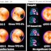


![(A-C) Representative whole-body maximum-intensity projection images and regional fused PET/CT images from three histologically confirmed osteosarcoma patients who underwent paired [68Ga]Ga-B7-H3-BCH PET/CT and 18F-FDGE PET/CT within seven days. (D) Multimodal imaging evaluation of Patient Three, including x-ray, MRI (T2-weighted imaging, T2WI), CT, and B7-H3 PET/CT.](https://img.auntminnie.com/mindful/smg/workspaces/default/uploads/2026/05/mei.XUQJWkpAJI.jpg?auto=format%2Ccompress&dpr=2&fit=crop&h=167&q=70&w=250)


