Radiologists from Johns Hopkins University applied cinematic rendering to the clinical management of congenital vascular anomalies and uncovered critical features of patient anatomy and pathology that were not evident using traditional volume rendering techniques or conventional CT.
Cinematic rendering employs a global lighting model to generate photorealistic 3D reconstructions of images. In recent years, several groups have demonstrated the potential of the advanced reconstruction technique to benefit the diagnostic evaluation of cancer and the examination of various internal organs and soft-tissue injuries and fractures.
This early work in cinematic rendering has shown the technique to be particularly useful in visualizing intricate, often highly vascularized anatomy, and it may help improve visualization of pediatric vascular anomalies, presenter Dr. Hannah Recht noted in a digital education exhibit at RSNA 2019.
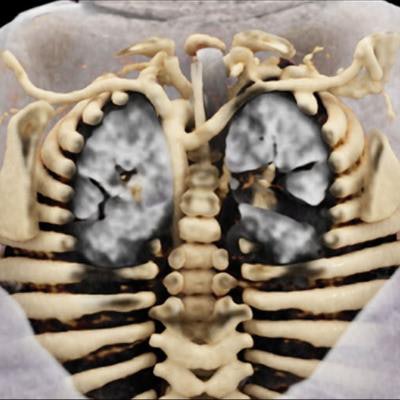
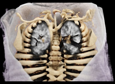
 The cinematic rendering shows a posterior view of an aberrant right subclavian artery coursing to the enteric tube, which is difficult to see using conventional CT and volume rendering. Image courtesy of Dr. Hannah Recht.
"Vascular anomalies in pediatric patients are challenging cases from an anatomic perspective, and we felt that cinematic rendering could be particularly helpful in these cases for treatment and surgical planning," she told AuntMinnie.com.
To that end, Recht and colleagues retrospectively applied cinematic rendering to a series of cases involving a variety of congenital vascular anomalies, including the following:
- Aortic coarctation. Cinematic rendering delivered a markedly enhanced view of severe coarctation in a previously healthy 4-year-old boy, compared with conventional CT and maximum intensity projection reconstructions. The advanced technique also allowed for visualization of both of the prominent internal mammary arteries and their relationship to surrounding structures, compared with only one of the arteries being visible on conventional imaging.
- Pulmonary sling. Axial and sagittal views of standard CT scans revealed the presence of pulmonary sling in an 8-year-old boy with a heart murmur. Applying cinematic rendering to these scans added a layer of depth and precision that more clearly demonstrated the complex relationships among vascular structures, which could be helpful in preoperative planning.
- Intralobar sequestration. Whereas CT angiography scans were able to detect abnormal arteries and veins in a newborn with a congenital pulmonary airway malformation in utero, cinematic rendering revealed the origin and complete course of these feeding and draining vessels in multiple planes. Identifying the precise positioning of these structures is crucial for surgical treatment planning.
- Double aortic arch. A 22-month-old girl had a double aortic arch confirmed on CT angiography scans and subsequent volume-rendered images, but these conventional techniques left the right subclavian artery obscured by the clavicle. In contrast, cinematic rendering provided a full view of the vascular ring and showed it completely encircling the endotracheal tubes -- information that would likely be helpful in planning a thoracotomy.
For the wide spectrum of pediatric vascular anomalies examined, cinematic rendering provided enhanced views of internal anatomy and precise information on the size, location, and relative depth of pathology not achievable with conventional CT or volume rendering, Recht noted.
"Given the relative ease of creating cinematic rendering images, we envision the technique being created and utilized in 'real-time' for patient care," she said. "We believe that this information is promising for surgical planning, when the knowledge of the location, dominance, and origin of the vessels and adjacent structures in question is essential."
Recht and colleagues in the radiology department currently use cinematic rendering to facilitate evaluation for 30 to 50 cases per week, with growing interest from referring clinicians. To promote more widespread use of the technique, they are preparing to release an educational software application that would allow users to examine chest anatomy through cinematically rendered images based on CT scans.


