CHICAGO -- CT imaging shows that engineered stone countertop workers are at risk of developing the lung disease silicosis, according to research presented December 2 at the RSNA meeting.
In her presentation, Sundus Lateef, MD, from the University of California in Los Angeles shared her team’s findings showing that these workers commonly present with atypical and advanced features of silicosis.
“This is a new and emerging epidemic, and we must increase awareness of this disease process so we can avoid delays in diagnosis and treatment for our patients,” Lateef said in an RSNA statement.
Silicosis is a form of pulmonary fibrosis, caused by breathing in tiny bits of silica. Silica is a mineral commonly found in sand, quartz, and other types of rock. This means that people working in the construction and mining areas are more prone to silica exposure. The researchers also noted that many workers in the countertop fabrication industry are Spanish-speaking Latino immigrants who are vulnerable to unsafe workplace conditions.
Lateef and colleagues sought to describe imaging findings of silicosis on CT in engineered stone countertop workers. They also correlated imaging findings with clinical severity as measured by pulmonary function tests and assessed awareness among primary providers and radiologists.
The pilot study included 55 patients at an urban safety-net hospital with few historic cases of silicosis, though the team included complete data from 21 patients for initial results. The patients, all of whom were Hispanic, were diagnosed with silicosis with available CT and pulmonary function tests.
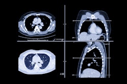
 Contrast-enhanced chest CT shows upper predominant fibrosis/lung scarring (red oval), solid masses adjacent to a left-sided pneumothorax (yellow oval) and diffuse silicotic nodules (purple circle). This patient had a history of working in the countertop-cutting industry (15 years), smoking cigarettes (10 pack years), and active tuberculosis (treated 7 years prior and excluded at the time of this image with laboratory testing). Image courtesy of the RSNA.
The team classified CT images as typical or atypical for chronic silicosis. Markers for atypical findings included mediastinal lymphadenopathy and upper-lobe predominant small nodularity and/or progressive massive fibrosis. All 21 patients were symptomatic, with the most common symptoms being dyspnea (n = 19) and cough (n = 17).
Both radiologists and primary clinicians showed low recognition of silicosis on first encounter. This included seven cases recognized by radiologists and four by primary clinicians. They suggested alternative diagnoses in most cases.
On secondary retrospective review, the team found that 11 cases were typical for silicosis, while the other 10 cases had atypical imaging features. Pulmonary function tests meanwhile showed a restrictive pattern in 18 cases.
Also, patients with consolidations had lower diffusing capacity for carbon monoxide (DLCO) test scores than patients without large opacities (18.1 vs. 24.5, p = 0.02).
However, the team pointed out that these findings “should be interpreted with caution prior to the analysis of all 55 patients.”
Lateef and colleagues are working on the ongoing California Artificial Stone and Silicosis (CASS) Project. The project’s goal is to promote respiratory health among vulnerable workers in California’s countertop fabrication industry.
For full 2024 RSNA coverage, visit our RADCast.


