AuntMinnie.com presents the 14th in a series of columns on the practice of ultrasound from Dr. Jason Birnholz, one of the pioneers of the modality.
Fellow Ultrasounder,
Belated happy new year, all, and may you also enjoy a prosperous Year of the Horse, which is my own Chinese birth sign. The end of a year is the arbitrary time when we think back on the best of the previous year and resolve to see that things are better as we restart with an almost clean slate.
 Dr. Jason Birnholz.
The start of December was also the time of the RSNA meeting, with its educational components dealing with "now" and research presentations hinting at "tomorrow." My first RSNA was in 1971 at the Palmer House. As a second-year resident in diagnostic radiology, I presented two papers, one of which was about uterine opacification during intravenous pyelography. The other was combining A- and B-mode ultrasound and the reflective properties of the front and back walls of structures to find a clear distinction between true cysts, solid masses, and semisolid masses like lymphomas and endometriomas.
This doesn't seem like much now, but it was a major concern in those pregrayscale, manual scan, "bistable" imaging days. Since then, I've missed only two meetings, one when the society moved it to Washington, DC, and another one when I was a visiting professor in Japan. I have also presented one or more papers as first or only author at 39 near-consecutive meetings, mostly but not entirely about ultrasound topics.
Fiduciary responsibilities
I am mentioning this bit of personal history because I work and go to RSNA and other meetings as a radiologist, not as an ultrasound monomaniac. And, because radiologists of my vintage always did something else first, I also think of myself as a diagnostic internist who uses imaging tools. That means that I, like other radiologists, have a primary responsibility to every patient I see that is identical to any other direct patient-physician interaction. Frank Chervenak has emphasized that patient care is a "fiduciary" relationship.
I need to be certain that the study I am about to perform is the safest and most effective way of providing relevant clinical information for what is known at the time of referral and of explaining to the patient what I perceive to be in his or her best interest. I also need to be certain that I am relating my findings and interpretations to the referring caregiver expeditiously, so that therapy can be started or modified or additional testing can be obtained without delay.
I am also responsible for the equipment that I use and how it is used. If I choose to work through an agent, i.e., a technologist, then I am responsible for the professional conduct and technical education and proficiency of that person, and for quality control in all facets of the exam. In a large department, those last sets of responsibilities may be shared by the section chief or department chairman.
Malpractice cases involving ultrasound exams typically name the technologist, attending physician, and supervisory physicians and their institutions, and there is often an allegation of old or inadequate equipment. In the case of teleradiology, the remote physician is responsible for interpretation of the images sent. If there is perceived negligence in any facet of acquiring images provided for interpretation, then the technologist and his or her supervisors and employer share the entire legal burden.
Some imaging centers are under the misapprehension that a referral or prescription is necessary for ultrasound. Patients can decide to come to me de novo, just as they can opt to go to or avoid any other (licensed) physician. I prefer that patients be referred, but it is the patient's choice that is central. In a framework of fiduciary responsibility, it is also fine for any physician to use ultrasound as a part of their care, provided that he or she has achieved at least a community standard of competence for the technique.
In the carefree mid-1980s, I often felt like a jungle guide, taking patients on a personal inner tour. I wonder if we really are not a lot more like pilots of river barges with priceless cargo crossing a torrent with lurking perils and barely visible whirlpools. Hemingway said something like, "It is the journey that matters, in the end." A good metaphor for what happens after we contact the probe to the skin, don't you think?
An exemplary image
As a radiologist who subspecializes in ultrasound, I have to be up-to-date on all the imaging alternatives for any potential referral. This past RSNA, with its emphasis on partnerships of all kinds for medical imaging, was also very unusual technically. I have never seen so many fascinating CT and MR images, or so few novel or interesting ultrasound examples.
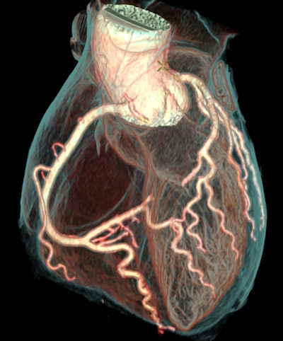
For example, the coronary artery definition and the simulation of a myofibrillar background texture on this high-speed CT image from a work-in-progress GE Healthcare Revolution scanner just blew me away.
 Image courtesy of Girish Muralidharan of GE Healthcare.
Skulking around various ultrasound booths, I saw technologists looking carefully at equipment and relatively smaller numbers of physicians and administrators, who seemed more concerned with finance issues than clinical performance. The salesmen, as usual, were touting high-end equipment with product-specific buzzwords, while the applications people I watched were usually displaying tablets or other small-screen devices, which were described as having the "same image quality" as a company's higher-end units. Indeed, the smaller the screen, the less noise will be visible, and the better the perceived contrast.
The CT and MR images utilized all kinds of computer graphics and image processing techniques, whereas ultrasound images were mostly those captured directly and without much in the way of postscan enhancement or data extraction. CT images, like x-rays, show atomic-level features, and MRI is at a molecular level of organization, while ultrasound is macroscopic or eye level for detail. But all three realms have very high information content for the things they depict. The question is: Where is ultrasound as useful, astonishing, or even just as esthetically pleasing as the CT image shown above?
The specialty morass
There is another factor that has had an effect on technically advancing ultrasound: its dispersion over many specialty areas. The earliest ultrasound pioneers were from all specialties, and there was a common patient-centered goal for our activities. Eventually and for a while, radiology provided a home for our nomadic technology.
Now we seem to be returning to our origins, but somehow without a transfer of the knowledge acquired during the 30 years or so of radiologic centralization. Every group that gets involved with ultrasound always seems to want to start from scratch. I think a lot of current authors of papers on point-of-care ultrasound would be astonished at what a thorough review of the older literature would reveal.
In a similar vein, I am both sad and happy when someone new to ultrasound has his or her rediscovery of the fabulous clinical value of ultrasound paraded in the popular press. Clinical dispersion while the method is still in a process of technical advancement also sends mixed messages to manufacturers who themselves have internal disputes between engineering and continued refinement and marketing, which seeks to make the most of what has already been produced.
 The ultrasound schooner passing glories into a storm. Image courtesy of Visual Jason Imagery.
Personal anecdotes don't mean much, but I have always been a little saddened by an experience many years ago when cross-cultural issues dominated. In the mid 1970s, when I was at the Peter Bent Brigham Hospital (now the Brigham and Women's Hospital), I got a lot of exercise trudging a 300-lb, foot-locker-sized Varian phased-array system from the ultrasound suite (actually a single subbasement cell, smaller than 8 x 10) to the coronary care unit.
The idea was to use high-speed imaging to look for and grade regional wall movement abnormalities. I was working with Josh Wynne, who was my junior faculty equivalent in cardiology then and who is now dean at one of our country's best medical schools. I presented our paper of observations of a reasonably sized group of patients at the 1979 American College of Cardiology (ACC) meeting. We showed what may have been the first videos (16-mm movies made from videotape so they could be projected on a big screen) of akinetic and dyskinetic segments of the left ventricle in patients with acute myocardial infarctions.
Was this the sensation of the meeting? Nope. The general reception was overtly hostile, and the comments of the session moderator, who had no personal or institutional experience with phased-array ultrasound, were dismissively negative. That kind of interspecialty distrust seems somehow to have been an unanticipated part of our postgraduate medical education system.
Specialty fragmentation is like a lysozyme that keeps ultrasound from gelling as a general multipurpose technique. Remember Abe Lincoln's comment about a house divided against itself? We need to centralize or cross-fertilize. Or, we can use new IT jargon and recognize the need to "break down the silos."
An evolving game plan?
Ultrasound seems to be buffeted on all sides. Having gotten to wherever we are now, most of us might opt to leave things alone. The pace of knowledge acquisition keeps accelerating, and global information access and exchange nears warp speed. So, we really cannot stand still operationally or intellectually.
At the acute-care, major hospital end, I think ultrasound should be limited pretty much to urgent uses in the emergency room, operating room, labor and delivery suite, and various intensive care units, with each area taking care of its own needs directly. We need to get away from piecemeal and excessive billing. When ultrasound needs to be done, it should be executed promptly and expertly and for the lowest practical fixed cost.
In compensation, there should be much more ambulatory outpatient ultrasound, particularly early diagnostic and screening protocols for everyone, not just designated risk populations. Ultrasound imaging needs to be available in any facility that sees patients routinely; it needs to be available when the patient is there, without prescheduling, and it should be integrated directly into each office visit unless there is a strong presumption that it cannot provide any useful information.
Ultimately, I would like to see ultrasound used as a primary physical exam tool for everything ultrasoundable. Physical exams remain a central component of pediatrics, perhaps primarily because of avoidance of invasive procedures or ionizing radiation exposure. The trend in adult internal medicine has been toward devaluing the traditional physical as insensitive and incompatible with the limited time commitment for patient-physician contact that has been occurring for ambulatory outpatient work.
The medical necessity of streamlining initial diagnoses and the need to avoid time-delaying imaging procedure scheduling are driving forces for this operational realignment; however, it will become practical because of a recent major advance with conventional array transducers that results in greatly improved penetration and sensitivity and the potential for quantitative and standardized ultrasound data acquisition. The improvement will be marked for small, portable systems.
Transparency
Second, as we are moving into an era of integrated electronic records, it will be easier than ever for ultrasound people to learn the outcome of their diagnostic efforts. Exact follow-up information is essential for learning and, at a unit level, for quality control. There are simple metrics such as the correspondence of ICD codes at the exam and later in the diagnostic process, the time to a final diagnosis, or the number of procedures required between initial ultrasound and case resolution that can be good ways of scoring clinical performance.
An algorithm has been developed for comparing surgical complication rates nationally: the National Veterans Affairs Surgical Quality Improvement Program (NSQIP); something much simpler will work for ultrasound. You might also be interested in a "Brief history of quality movement in U.S. healthcare." Establishing performance standards needs to be done nationally, and the metrics need to consider wellness, not just diagnoses of diseases or disease states.
News and next time
There was a recent announcement on Phys.org of a new finding about ultrasound wave propagation from three Dutch universities. The bursting of insonified nanodroplets of perfluorocarbon (which are very fragile at body temperature) was captured with an optical camera recording at 25 million frames per second. The phenomenon being observed is a form of destructive energy concentration by relatively large ultrasound pulses on targets that are nanometer-scale sizes.
The new theory indicates that very high-frequency harmonics are generated by velocity dispersion during pulse propagation. I wondered if a similar mechanism might not be involved in how Doppler pulses generate shear waves. The article goes on to mention new possibilities for selective chemotherapy by lysing drug-filled nanodroplets when they become concentrated within the vascular bed of a tumor, without harming adjacent normal tissues.
The main reason I am featuring this exemplary bit of basic research is that after more than 50 years of an enormous amount of research, here is something new about ultrasound pulse propagation in the body. From time to time, we need to remind ourselves that ultrasound propagation in tissues is an extraordinarily complex (or ill-behaved) process, much more so than is the case with x-ray photons or magnetic fields. It is to our credit that we get as much out of ultrasound as we do. It is also sobering to remember that as safe as we know ultrasound to be, we still do not know everything about the way ultrasound interacts at a cellular or subcellular level.
In my next article, I am going to return to actually doing ultrasound and will show you something that is new, visually spectacular, and available to all.
Dr. Jason Birnholz was one of the few advanced academic fellows of the James Picker Foundation, and he has been a professor of radiology and obstetrics. He is a fellow of the American College of Radiology and the Royal College of Radiology, and he was an associate fellow of the American College of Obstetricians and Gynecologists.
The comments and observations expressed herein do not necessarily reflect the opinions of AuntMinnie.com.


