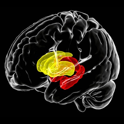
One of the largest-ever neuroimaging studies of individuals with HIV used MRI to reveal that lower current CD4+ T-cell counts were associated with smaller hippocampal and thalamic volumes in the brain. Also, the subset of participants not receiving treatment had smaller putamen volumes, according to findings published January 15 in JAMA Network Open.
Furthermore, detectable HIV load was associated with smaller hippocampal volumes across all study participants, but in the subset of participants receiving HIV treatment, detectable viral load was also associated with smaller amygdala volumes.
A research team led by Talia Nir, PhD, a postdoctoral scholar at the University of Southern California (USC) Stevens INI's Laboratory of Brain eScience (LoBeS), and colleagues pooled MRI data from 1,203 HIV-positive individuals across Africa, Asia, Australia, Europe, and North America.
They extracted volume estimates for eight subcortical brain regions from T1-weighted MR images to identify associations with blood plasma markers of current immunosuppression (CD4+ T-cell counts) or detectable plasma viral load in HIV-positive participants.
Nir and colleagues found lower current CD4+ cell counts were associated with smaller volumes in the hippocampus and thalamus volumes. Taking immunosuppressant drugs was also associated with larger ventricles.
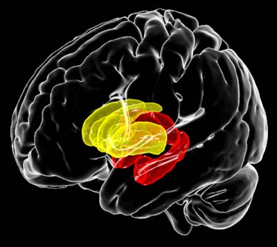
 The shift of HIV infection from a fatal to chronic condition in the era of more widely available treatment appears to be accompanied by a shift in the profile of HIV-related brain abnormalities beyond the basal ganglia (yellow), frequently implicated in earlier studies, to limbic structures (red). MR image courtesy of Talia Nir, PhD; Neda Jahanshad, PhD; and James Stanis of the Mark and Mary Stevens Neuroimaging and Informatics Institute at the Keck School of Medicine of USC.
However, study participants that didn't take combination antiretroviral therapy had lower CD4+ cell counts and smaller putamen volumes. A detectable plasma viral load was associated with smaller hippocampal volumes. The detectable plasma viral load was also associated with smaller amygdala volumes in patients receiving combination antiretroviral therapy.
"Through these large-scale efforts, we're beginning to understand the link between immune function and brain alterations in individuals living, and aging, with HIV," the study authors said.
Next, the researchers will analyze imaging data over time, including diffusion imaging data, to further understand how clinical markers of HIV infection affect the brain and the rate of neurodegeneration.


