Researchers from Paris have provided comprehensive new details about the methods and techniques used to obtain images of the human brain in vivo on an 11.7 tesla (11.7T) MRI system.
"The safety and imaging data gathered in this first study confirm that humans can safely tolerate such intense magnetic fields and that brain images at mesoscale resolutions can be achieved in a reasonable time," noted Prof. Denis Le Bihan, PhD, and Nicolas Boulant, PhD, both from the NeuroSpin research facility and the Iseult Project of the French Alternative Energies and Atomic Energy Commission (CEA), in an accompanying editorial published in Nature Methods on 17 October.
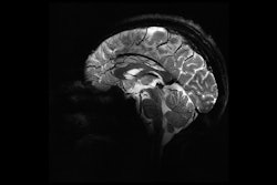
 In vivo images of the human brain obtained at 11.7T. (A) In vivo 3D T2 variable flip angle turbo spin-echo acquisition at 11.7T with parallel transmission GRAPE universal pulses (resolution 0.55 × 0.55 × 0.55 mm3, acquisition time 13 min) demonstrating the high B1+ (RF) field homogeneity reached throughout the whole brain volume. (B) In vivo T2*-weighted 2D GRE sagittal slice (resolution 0.2 × 0.2 × 1 mm3, acquisition time 8 min 30 s). (C) T2* weighted 2D GRE axial images acquired at 3T (left), 7T (middle) and 11.7T (right) with identical acquisition times (4 min 17 s), while keeping similar contrast-to-noise ratio through adjusted acquisition parameters (FA (°), TR (ms) and TE (ms) of 27, 750 and 45; 34, 950 and 25; 27, 600 and 20 at 3, 7 and 11.7T, respectively) and spatial resolution (0.5, 0.325 and 0.2-mm in-plane resolution for 3, 7 and 11.7T, respectively, 1-mm thickness). (D) The 11.7T T2*-weighted 2D GRE axial images (resolution 0.19 × 0.19 × 1 mm3, acquisition time 5 min 16 s) juxtaposed to turbo spin-echo T2-weighted images (resolution 0.3 × 0.3 × 1 mm3, acquisition time 4 min 26 s). All figures courtesy of Nicolas Boulant, PhD, et al and Nature Methods.
Before the study began, the team performed tests with human blood samples and on animals in vivo to determine the impact of an ultrahigh field (UHF). Post authorization for scanning human subjects, protocols involving physiological, cognitive, and genotoxicity tests were conducted to verify the safety of large magnetic fields for humans.
The first in vivo exploratory study took place in 2023, testing 20 people scanned at 11.7 tesla and another cohort of 20 people with the magnetic field switched off to obviate potential psychological bias, with each participant believing that the magnetic field was on for the scan. The study showed that no significant differences between the two groups were apparent in any of the biological tests. There were no statistically significant differences in micronuclei count for genotoxicity for each participant before versus after 1 hour and 30 minutes of exposure at 11.7T.
Follow-up phone calls by the NeuroSpin medical team up to a week after exposure at 11.7T did not indicate any abnormality. Six and four out of 20 volunteers exposed to the 0 and 11.7T field, respectively, reported fatigue the same day or the day after. Four volunteers from the 0-tesla field group and one from the 11.7T group reported headaches. Subjects from the groups also experienced transient dizziness when entering and exiting the magnet, as well as a metallic taste in the mouth, a known minor side effect of MRI.
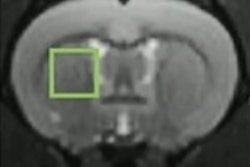
 T2*-weighted 2D GRE axial images acquired at 3T, 7T, and 11.7T (different subjects). Acquisitions were performed with resolution = 0.2 × 0.2 × 1 mm3, FA = 27°, TE = 20 ms, TR = 0.6 s, bandwidth = 40 Hz/pixel, acquisition time = 4 min 20 s.
First author Nicolas Boulant, PhD, head of the Iseult Project and director of research at the CEA, and colleagues were also satisfied with the quality of the images acquired on the first volunteers, which exceeded the team's most optimistic expectations, the authors noted in the media. They acquired structural in vivo images of the human brain with a high resolution down to 0.19 x 0.19 x 1 mm3 in around five minutes and with good signal uniformity without serious artifacts of RF field inhomogeneity. Furthermore, these images indicated sufficient SNR to boost spatial resolution further, they added.
Future steps and follow-up
The study has proved that humans can be scanned safely with powerful magnetic fields, while brain images of mesoscale resolution can be obtained within a reasonable time frame, according to the authors. This tool will allow researchers to understand aging and neurocognitive disorders and provide them with new opportunities to better understand specific neurological and psychiatric disorders, as well as help to develop new disease biomarkers and therapeutic tools, across diseases such as drug-resistant focal epilepsy, multiple sclerosis, and neurodegenerative diseases such as Alzheimer's and Parkinson's, they explained.
The study was exploratory and it was not possible to examine multiple types of MRI image contrast, in particular functional MRI (fMRI) and diffusion tensor imaging. The group also noted that around two-thirds of the high-resolution scans were corrupted by motion. Future studies will focus on the development and implementation of motion compensation tools, as well as highly accelerated image acquisition sequences.
Future studies will also include the use of more efficient RF coils and more powerful gradients to further increase performance for high-resolution functional MRI and diffusion tensor imaging or tractography to map brain connectivity and study the structural-functional relationships that make up the human brain, which could lead to a better understanding of the biological mechanisms underlying mental disorders and consciousness.
"At this stage, we are considering writing a complete article showing the safety of UHF MRI (we have more extensive results beyond those shown in the Brief Communication Nature Methods article) and getting administrative approval to scan subjects and patients, beyond the initial cohort of 40 subjects," corresponding author Le Bihan told AuntMinnie.com on October 21. "We will aim first at getting high-resolution fMRI data."
You can read the full paper here.