
The clinical benefits of photon-counting CT (PCCT) are becoming more and more clear, according to a presentation delivered September 6 at the International Society of Computed Tomography (ISCT) 2023 meeting in San Diego.
Because photon-counting detectors directly record individual photons and their associated energy, the technology offers multiple benefits over CT imaging using conventional energy-integrating detectors, said presenter Cynthia McCollough, PhD, of the Mayo Clinic in Rochester, MN.
"Clinically [PCCT allows us to see] things better and see smaller things ... with less radiation and contrast," she told session attendees.
Seeing small structures better
McCollough began her presentation with an overview of the benefits of PCCT, which include the following:
- An ability to exclude electronic noise
- Increased iodine contrast-to-noise ratio and dose efficiency
- Increased iodine at high kV
- Reduced beam hardening and metal artifacts
- Improved special resolution
- Lower radiation dose
- Simultaneous, single kV, multi-energy CT imaging
- Multi-energy ultrahigh resolution and cardiac CT
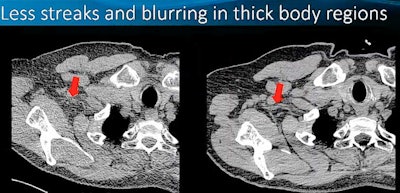
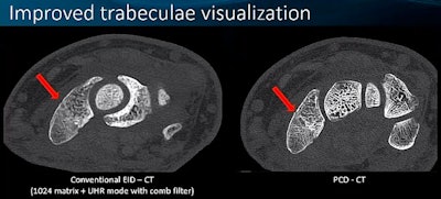
She then "unpacked" some of these benefits, noting that PCCT not only produces images of thick body regions with less streaks and blurring and clearer images in patients with metal artifacts such as spine hardware compared to conventional CT. It also allows clinicians to see smaller things (for example, the anterior choroidal artery), improves visualization of the trabeculae, and detects small calcifications that conventional CT misses.
 All images courtesy of Cynthia McCollough, PhD, and Mayo Clinic.
"The photon-counting detector process allows us to get a consistent iodine signal," McCollough noted. "What does that do? It gives us brighter iodine, and since more than half of our exams use iodine, that's a really big deal."
Seeing new structures
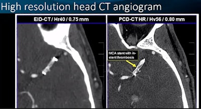
Not only does the technology visualize smaller body structures compared with conventional CT imaging but it also visualizes new things, according to McCollough. She presented a conventional image of a head CT angiogram and a PCCT version; the PCCT image clearly showed an MCA stent with in-stent thrombosis, as well as two images (one conventional, and one PCCT) of short and long posterior ciliary arteries.
"[Our peers say that they're seeing] ciliary arteries they haven't seen before," McCollough said.
The technology reduces image noise at the same radiation dose, she explained, and it offers simultaneous high spatial resolution and multi-energy imaging.
Finally, McCollough stressed that PCCT imaging can improve patient management, for example by lowering Coronary Artery Disease Reporting and Data System (CAD-RADS) scores for patients with dense calcifications and identifying cerebrospinal fluid leaks more efficiently.
"This is the kind of patient life-changing care that we [can offer] because we have better spatial resolution and energy information," McCollough concluded.


