Photon-counting CT (PCCT) technology can reduce the dose on CT lung cancer screening exams to the equivalent of chest x-rays, researchers have reported.
"[Our study found that] semiautomated nodule detection is highly sensitive in [PCCT] x-ray dose scans," wrote a team led by Bjarne Kerber, MD, of the University Hospital Zürich. The study results were published March 10 in Investigative Radiology.
Reducing radiation dose on CT imaging is key to providing patients with the best care, and PCCT has been shown to help do that. Using PCCT for lung cancer screening shows promise in this regard.
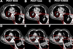
For their study, Kerber's group evaluated the effect of PCCT dose reduction to x-ray equivalent levels on lung imaging nodule detection, diameter, volume, and density compared to a low-dose reference standard using semiautomated and manual methods. They conducted a study that included 101 patients who underwent same-study noncontrast low- and chest x-ray-dose CT scans with PCCT imaging between February and July 2023. Nodule detection and measurement of their diameters and volumes was performed semiautomatically using computer-aided diagnosis software; two radiologists manually measured nodule diameters and nodule density, then classified the nodules using Lung-RADS v2022.
The team reported the following:
- Mean volume CT dose index for x-ray dose scans was 0.11 mGy compared with 0.65 mGy low-dose images (p < 0.001).
- The semiautomated method showed high overall sensitivity for nodule detection (94%), with a higher sensitivity for solid nodules (95%) and lower sensitivity for subsolid nodules (86%).
- Semiautomated measurements underestimated nodule diameter for solid nodules on x-ray dose scans (p = 0.01), but the researchers did not find a significant effect for nodule volume (p = 0.775).
- The two radiologist readers rated nodule density as less dense on x-ray dose compared with low-dose scans.
- The study found no significant difference in nodule diameter between radiologist readers between scan doses, and there were good to excellent correlations between semiautomated and reader nodule diameter measurements.
- Agreement and accuracy between low-dose and x-ray dose Lung-RADS classifications across methods were good, at kappa 0.73 for low-dose and 0.62 for x-ray dose.
The takeaway? "Accurate semiautomated and manual nodule measurements are feasible on x-ray dose scans, but nodule density [tended to be] underestimated," the group concluded.
The complete study can be found here.