An MRI-based radiomics model shows potential for distinguishing low- from high-risk cases of ductal carcinoma in situ (DCIS), an early form of breast cancer, according to a study published April 1 in Radiology.
The study highlights how radiomics can help stratify risk in DCIS cases compared with clinical and qualitative imaging information alone, noted lead authors Kalina Slavkova, PhD, of Columbia University in New York City, and Ruya Kang, PhD, of Brown University in Providence, RI.
“Current breast MRI interpretation approaches are sensitive to ductal carcinoma in situ (DCIS) detection but are not able to identify DCIS at risk for upstaging, which may lead to overtreatment of low-risk participants when surgery is pursued in lieu of active surveillance,” the group wrote.
About 20% of new breast cancers in the U.S. are diagnosed as DCIS, an early form of breast cancer that forms in the milk ducts, based on dynamic contrast-enhanced (DCE) MRI and biopsies. Most women undergo surgery, yet only 26% of DCIS cases are subsequently identified as invasive disease, the authors noted.
Hence, the researchers developed a clinical and DCE MRI-based radiomics model and evaluated it for predicting low- and high-risk DCIS cases. In a secondary outcome, they compared the model to a genetic test (Oncotype DX Breast DCIS Score), which determines risk (low, intermediate, or high) using specimens obtained during surgery.
 A graphical abstract of the study. Image courtesy of RSNA.
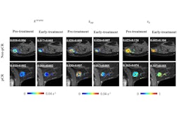
The group culled data from 297 women with DCIS with median age of 60 years old. They used the publicly available Cancer Phenomics Toolkit, or CaPTk, software to compute 65 radiomic features, with participants subsequently clustered into risk groups based on their radiomic features. From these, they identified radiomic phenotypes that were positively associated with upstaging to invasive disease following surgery.
According to the analysis, combining the top three radiomic principal components with clinical and qualitative MRI factors enabled the classification of 24.7% more participants in whom disease did not upstage, and hence were considered low risk and safe for active surveillance.
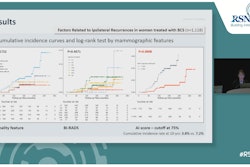
“We found that radiomic phenotype assignment was positively associated with upstaging to invasive disease (p = 0.007), suggesting that the identified phenotypes may correspond to low- and high-risk groups,” the group wrote.
However, the radiomic phenotypes and principal components were not associated with the Oncotype DX Breast DCIS Score, they noted.
Ultimately, further research to identify additional biomarkers that can help improve prediction of low-risk cases, such as novel pathologic features, should be explored, the group suggested.
In an accompanying editorial, Soo-Yeon Kim, MD, and Ok Hee Woo, MD, breast imaging experts at Korea University Guro Hospital in Seoul, noted that the study was well designed and effectively highlights the potential of MRI-based radiomics in predicting the invasive upstaging of DCIS.
“We look forward to future research on this clinically significant topic,” they wrote.
Work to further improve risk stratification for DCIS may include advancing segmentation techniques, incorporating delayed phases in DCE MRI, exploring the full potential of basic qualitative MRI features, and integrating mammographic and MRI characteristics, Kim and Woo concluded.
The full study can be found here.