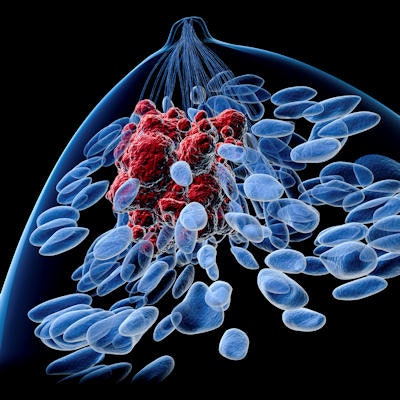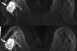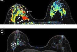
A new type of diffusion-weighted imaging (DWI) is giving conventional breast MRI a run for its money. Field-of-view optimized and constrained undistorted single-shot (FOCUS) imaging can identify 85% of suspicious breast lesions, ECR 2018 delegates learned in Vienna.
Researchers have investigated DWI as a method to overcome the limitations of conventional breast MRI. In particular, they've looked at the detection and characterization of breast cancer with the aim of improving the diagnostic performance of dynamic contrast MRI, said Dr. Lorenzo Vassallo from the radiology department at Istituto di Candiolo-IRCCS-Fondazione Piemontese per la Ricerca sul Cancro Onlus in Candiolo, Italy.
However, because of the variability of acquisition and analysis methodologies at all the different centers, there is not a set value for the differentiation between benign and malignant breast lesions. In addition, the spatial resolution of standard DWI can affect the evaluation of morphological descriptors of the breast lesions, he added.
"This new sequence will provide high-resolution images with more accurate characterization of masses, significant reduction of architectural distortion, less artifacts, and equivalent evaluation," he said.
To test this hypothesis, he and his colleagues compared the visibility of breast lesions at 1.5 tesla (Optima 450w GEM suite, GE Healthcare) using FOCUS DWI versus standard DWI and dynamic contrast-enhanced (DCE) MRI sequences. They enrolled 50 consecutive women undergoing presurgical breast MRI from January to December 2016.
They performed FOCUS DWI on the axial plane with limited shimming to the rectangular area target. DCE MRI was considered the reference standard. The type of each suspicious lesion detected at DCE MRI was evaluated and then compared with similar findings identified at both standard DWI and FOCUS DWI. The researchers rated the lesions as +1, meaning the lesion had better visibility on FOCUS; -1, the lesion had better visibility on standard DWI; or 0, neither was better. The same analysis was performed for FOCUS and DCE MRI.
DCE MRI identified 59 malignant, pathologically proven lesions: 48 mass and 11 nonmass lesions. All of the lesions were correctly detected at standard DWI. Five lesions -- all masses ranging in size from 7 mm to 15 mm -- were not identified at FOCUS DWI.
| Comparison of FOCUS DWI and standard DWI in Italy | |||
| No. of mass lesions (%) | No. of nonmass lesions (%) | Total (%) | |
| -1 | 12 (28%) | 7 (64%) | 19 (35%) |
| 0 | 14 (32%) | 1 (9%) | 15 (28%) |
| +1 | 17 (40%) | 3 (27%) | 20 (37%) |
| Total | 43 (100%) | 11 (100%) | 54 (100%) |
| Comparison of FOCUS DWI and DCE MRI in Italy | |||
| No. of mass lesions (%) | No. of nonmass lesions (%) | Total (%) | |
| -1 | 33 (77%) | 11 (100%) | 44 (81%) |
| 0 | 10 (23%) | 0 | 10 (19%) |
| +1 | 0 | 0 | 0 |
| Total | 43 (100%) | 11 (100%) | 54 (100%) |
"When asking readers to express a preference in terms of visibility between FOCUS and standard DWI, in the majority of cases, the visibility was considered +1 or even better at FOCUS compared with standard -- and in particular for mass lesions compared with nonmass lesions," he said.
However, when comparing FOCUS DWI with DCE MRI, the visibility of the lesions was never better on FOCUS DWI, although it was comparable in 10 cases of mass lesions.
The results suggest a future for FOCUS DWI in the evaluation of breast lesion morphology, but multicenter studies are required to confirm the preliminary results in a larger series of patients, Vassallo said.


