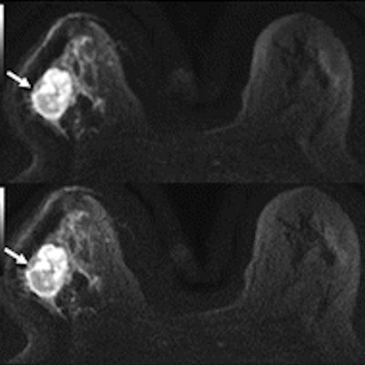
A promising new protocol for diffusion-weighted MRI (DWI-MRI) resulted in better image quality and improved signal-to-noise ratio for breast lesions, according to a prospective study published online on May 29 in Radiology: Imaging Cancer.
The protocol, multiplexed sensitivity-encoding (MUSE) DWI, splits a single-shot imaging acquisition into multiple shots, which can help reduce the technique's sensitivity to motion. Researchers from the University of Arizona had previously explored MUSE-DWI's potential to produce high-quality brain images with a 15-minute MRI scan, but its use for breast imaging hasn't been as readily studied.
"MUSE DWI yielded significantly improved image quality compared with single-shot DWI in phantoms and participants," wrote the international team of authors, led by Dr. Isaac Daimiel Naranjo from the breast imaging service at Memorial Sloan Kettering Cancer Center in New York City.
DWI analyzes the distribution of water to evaluate breast tissue structures at a microscopic level without the need for a contrast agent. Prior research has found the MRI technique can reduce false-positive findings and increase breast cancer detection.
However, single-shot DWI can also lack high spatial resolution and is especially sensitive to patient motion. The authors investigated the MUSE protocol for DWI as one solution to overcome these limitations, while also potentially speeding up contrast-free MRI times.
For their prospective study, the authors optimized MUSE DWI for breast phantoms. They tested multiple MUSE DWI acquisition techniques and selected one with the best quality for breast imaging.
Once the protocol was established, the authors enrolled 30 women with BI-RADS category 2-5 lesions found through contrast-enhanced MRI. The patients first underwent imaging with DWI, followed immediately by MUSE DWI.
Two breast imaging radiologists independently read the DWI and MUSE DWI images, grading each images' fat suppressing, artifacts, lesion visibility, and overall quality.
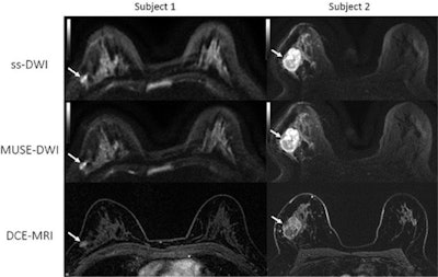
 Comparison of single-shot DWI (ss-DWI), MUSE-DWI, and dynamic contrasted-enhanced (DCE-MRI) axial images for two patients with biopsy-proven invasive ductal carcinoma. Image courtesy of the RSNA.
The researchers consistently ranked the quality of MUSE DWI images higher than the quality of single-shot DWI. One author rated MUSE DWI images higher in 58% of cases, while the other ranked MUSE DWI higher 65% of the time.
"In accordance with these previous investigations, our preliminary study in the breast found that the quality of the image with MUSE DWI was ostensibly better, resulting in improved lesion delineation compared with single-shot DWI," the authors wrote.
Further, MUSE DWI demonstrated significantly better signal-to-noise ratio and improved fat suppression and artifact correction. The researchers also found no difference in the apparent diffusion coefficient values of the two procedures.
Because the MUSE technique improved DWI image quality, the authors noted it may be possible to cut down on MRI scan time while keeping image quality consistent. However, this study only tested the protocol with a small number of patients and a limited number of small lesions.
Although the findings look promising for clinical application, future studies should evaluate the protocol for smaller lesions, in shorter scan times, and compared with other DWI protocols, the authors noted.


