A new gallium-68 (Ga-68)-labeled PET/CT radiotracer appears superior over F-18 FDG for detecting bone metastases, particularly in patients with osteoblastic lesions and prostate cancer, a group in China has reported.
The finding is from a prospective trial comparing Ga-68 DOTA-IBA-PET/CT to F-18 FDG-PET/CT in 46 patients and supports the use of the tracer as a theranostics imaging agent, noted corresponding author Yue Chen, MD, of Southwest Medical University in Sichuan, and colleagues.
“Our study supports the rationale for using Ga-68 DOTA-IBA to improve the management of patients with bone metastases, considering the therapeutic efficacy of [lutetium-177] DOTA-IBA,” the group wrote. The study was published April 14 in Scientific Reports.
Theranostics is an innovative nuclear medicine treatment approach that combines diagnostic and therapeutic capabilities in a single agent. In a previous study, the group tested such an agent (DOTA-IBA) combined with Ga-68 to image bone metastases and with Lu-177 to treat bone lesions, with promising results in 18 patients.
In this study, the researchers further evaluated the Ga-68 DOTA-IBA tracer in a prospective trial, with a comparison to F-18 FDG, which is has proven good performance in detecting bone metastases in different cancers.
Between October 2022 and September 2023, the group enrolled 46 patients with confirmed primary cancer and with clear indications for F-18 FDG-PET/CT scans for initial staging or restaging after therapy. Twenty-two patients had lung cancer and 12 prostate cancer. Participants underwent F-18 FDG-PET/CT and then Ga-68 DOTA-IBA within an average of 3.1 days.
Three board-certified nuclear medicine physicians with more than five years of experience analyzed the images and classified lesions into five groups based on anatomical location (limbs, vertebrae, pelvis, ribs, and skull) and then compared lesion detection rates between the two techniques.
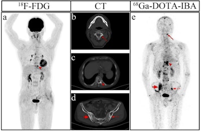
 Images of a 69-year-old man diagnosed with prostate cancer who had a history of endocrine therapy for two years. (a) The maximum intensity projection (MIP) image of F-18 FDG showed moderate radiotracer uptake in a thoracic vertebra. (e) The MIP image of Ga-68 DOTA-IBA demonstrated more lesions in the cervical vertebra and pelvis. (b) An osteoblastic lesion in the C3 vertebra right attachment (curved arrows) was negative on F-18 FDG (SUVmax = 2.0) and positive on Ga-68 DOTA-IBA (SUVmax = 8.7). (c) Another osteoblastic lesion in the T10 vertebra left side (arrowheads) was both positive on F-18 FDG (SUVmax = 5.2) and positive on Ga-68 DOTA-IBA (SUVmax = 35.0). (d) Osteoblastic lesions in bilateral ilium (dotted arrows and arrows) were negative on F-18 FDG (left: SUVmax = 1.4, right: SUVmax = 1.6) and positive on Ga-68 DOTA-IBA (left: SUVmax = 7.6, right: SUVmax = 8.7). Follow-up imaging three months later confirmed the presence of these bone metastases. Image and caption available for republishing under Creative Commons license (CC BY 4.0 DEED, Attribution 4.0 International) and courtesy of Scientific Reports.
Results indicated that Ga-68 DOTA-IBA had higher diagnostic efficacy than F-18 FDG in detecting bone metastases in the limbs (73.2% vs. 64.1%), vertebras (78.1% vs. 67.4%), ribs (86.6% vs. 62.2%), pelvis (78.6% vs. 68.9%), and skulls (80% vs. 38%).
For osteoblastic lesions, the detection rate for Ga-68 DOTA-IBA was 83.3% and for F-18 FDG, 51.5% (p < 0.001). In participants with metastatic prostate cancer lesions, the detection rate of Ga-68 DOTA-IBA was 84.7% and for F-18 FDG, 55% (p < 0.001).
“Ga-68 DOTA-IBA-PET/CT demonstrates superior diagnostic performance over F-18 FDG-PET/CT in detecting bone metastases, particularly in osteoblastic lesions and prostate cancer cases,” the researchers wrote.
Ultimately, early and precise diagnosis of bone metastases is crucial for patient management, according to the group. They noted a rich field of other radiotracers in the field, either approved for use or in development, such as F-18 sodium fluoride (F-18 NaF), Ga-68-labeled fibroblast activation protein inhibitor (Ga-68 FAPI), and Ga-68 prostate-specific membrane antigen (Ga-68 PSMA).
“There is a need to investigate Ga-68 DOTA-IBA versus these emerging imaging agents in the future as necessary,” the group concluded.
The full study can be found here.


