Researchers in Germany have proposed a new approach derived from brain PET imaging for diagnosing and staging Alzheimer’s disease, according to a study published March 25 in Radiology.
The method is based on the amount of the brain “filled” with three hallmark pathologic abnormalities associated with the disease – amyloid, tau, and neurodegeneration – and could become a valuable tool for clinicians, noted first author Elena Doering, MD, of the University of Cologne, and colleagues.
“Spatial extent-based amyloid-tau-neurodegeneration considerations, such as the amount of the brain 'filled' with pathology ('fill states'), may provide additional information for monitoring AD progression and screening at-risk individuals,” the group wrote.
Diagnosing and monitoring Alzheimer’s disease typically uses average standardized uptake value (SUV) ratios of PET radiotracers in regions known to be affected by the disease, the authors explained. Yet average SUV ratios largely ignore the spatial distribution of pathology, which can vary among individuals. Some patients with widespread pathology may exhibit more severe cognitive impairment compared with individuals with the same quantity of pathology confined to fewer brain regions, for instance.
The researchers suggested fill states as a new biomarker, and in this study, they tested the efficacy of fill states versus standard SUV ratios for reflecting stages of the disease, from subjective cognitive decline to dementia.
For data, the group used PET imaging from two cohorts, the Alzheimer’s Disease Neuroimaging Initiative (ADNI, n = 324) and the Tau Propagation over Time study (T-POT, n = 99). They computed fill states for amyloid, tau, and neurodegeneration using percentages of significantly abnormal voxels, the smallest unit in a PET image.
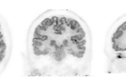
 Schematic illustrates how fill states are derived. (A) Derivation of an exemplary tau fill state. Note that different meta-regions of interest (ROIs) were recommended for the assessment of amyloid PET markers from carbon-11 Pittsburgh compound B (C-11 PiB) and F-18 florbetapir The bottom beige box provides definitions for the two distinct concepts of fill states and standardized uptake value (SUV) ratios. (B) Surface plots corresponding to exemplary fill states for amyloid, tau, and neurodegeneration. The gray-shaded area indicates the meta-ROI, and the colored area indicates abnormal voxels. Color coding: blue = amyloid, purple = tau, red = neurodegeneration. Con = control. Image and caption courtesy of RSNA.
According to the analysis, higher fill states were associated with higher stages of cognitive impairment (p < 0.001), and tau and neurodegeneration fill states showed higher diagnostic performance for cognitive impairment compared with SUV ratio (p < 0.05) across cohorts, the authors reported.
Similarly, all fill states were negatively correlated with cognitive performance (p < 0.001) and uniquely characterized the degree of cognitive impairment even after adjusting for SUV ratio (p < 0.05), the researchers noted.
“Fill states, representing the spatial extent of pathology, may serve as promising new monitoring biomarkers for Alzheimer disease, which reflect disease progression better than reference-standard standardized uptake value ratios,” the group wrote.
One potential application of fill-state data could be to serve as a readout for clinical trials, pending proof of longitudinal efficacy, the researchers added.
In an accompanying editorial, Mijin Yun, MD, PhD, and doctoral student Daesung Kim, of Yonsei University in Seoul, South Korea, wrote that the results are compelling and noted that the clinical efficacy of SUV ratios in quantifying PET scans has been repeatedly questioned, despite its widespread use.
"By shifting the focus from intensity-based quantification to spatial distribution, fill states could enhance early detection and diagnostic accuracy of [Alzheimer’s disease] progression," they wrote.
However, before fill states can be integrated into clinical practice, further research is needed to ensure the robustness of the model across imaging sites and PET radiotracers, as well as to validate their longitudinal stability.
“If these challenges are successfully addressed, fill states may become a valuable tool for the diagnosis and staging of AD in PET imaging,” Yun and Kim concluded.
The full study is available here.