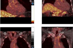
FDG-PET/CT scans have shown that vascular inflammation may be a trigger for high rates of heart attack and other adverse events in patients with pneumonia, according to a study published online December 14 in JACC: Cardiovascular Imaging.
A group led by Dr. Vicente Corrales-Medina of the University of Ottawa in Ontario, Canada, analyzed F-18 FDG scans in patients before and after they recovered from community acquired pneumonia (CAP), and found that higher aortic inflammation seen on the scans could be a sign for poor outcomes in these patients.
"Our findings may suggest a mechanistic link between vascular inflammation and [major adverse cardiovascular events]," the group wrote.
Patients with community-acquired pneumonia are at increased risk of major adverse cardiovascular events (MACE), and this risk remains elevated for months to years after the infection has resolved. Vascular inflammation in the aortic walls, as measured by F-18 FDG PET/CT scans, is strongly associated with the risk of MACE. Thus, increased vascular inflammatory activity post-community-acquired pneumonia could be a plausible mechanism for the elevated MACE risk in CAP survivors, the authors hypothesized.
"Yet, any putative changes in vascular inflammatory activity after CAP have never been examined," they wrote.
To fill the knowledge gap, the researchers enrolled 25 consecutive community-dwelling adults 65 years of age or older admitted to the Ottawa Hospital in Ontario between August 2015 and January 2018 with symptoms consistent with pneumonia of less than two weeks in duration. Study participants underwent F-18 FDG-PET/CT scans to detect aortic inflammation within the first 72 hours of their hospitalization and again 30 to 45 days after their hospital discharge.
Aortic inflammation in the pneumonia patients was significantly higher during the first 72 hours than after the patients recovered (maximum tissue-to-background ratio: 2.56 vs. 2.09, p < 0.001), the researchers found.
Moreover, when compared with 28 patients without pneumonia who had undergone FDG-PET/CT scans for suspected cancer (but who ultimately received a benign diagnosis), aortic inflammation in patients both during CAP and after recovery was also significantly higher, the group reported.
F-18 FDG-PET/CT has independent and incremental prognostic value in predicting MACE and the temporal pattern of F-18 FDG-PET/CT changes after CAP seen in the study correlated well with the trajectory of MACE risk in CAP survivors, the researchers added. But they noted that this was the first study to assess aortic F-18 FDG-PET/CT in patients with CAP and that additional research is warranted.
"Our findings may suggest a mechanistic link between vascular inflammation and MACE post CAP that needs to be further investigated," the group concluded.


