A new SPECT/CT radiotracer produces high quality and readily interpretable images of cardiac amyloidosis, a study presented at the 2024 Society of Nuclear Medicine and Molecular Imaging (SNMMI) meeting in Toronto has found.
A team led by Jonathan Wall, PhD, of the University of Tennessee Graduate School of Medicine in Knoxville reported that an amyloid-specific and pan-amyloid binding radiotracer designed for planar and SPECT/CT imaging -- called Tc-99m p5+14 -- produced clinically effective images that were interpretable at both one and three hours after injection.
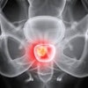
 Representative whole-body planar images of Tc-99m p5+14 of healthy subjects and, with SPECT/CT images, of a patient with ATTRv cardiac amyloidosis at 1 hour post injection showing no uptake in the heart of the healthy subject and intense signal in the heart of the patient using both planar and SPECT/CT imaging. Image and caption courtesy of the SNMMI.
"Patients with amyloid cardiomyopathy had significant Tc-99m p5+14 uptake in the heart, whereas no cardiac uptake was observed in healthy subjects," the group wrote.
Cardiac amyloidosis -- sometimes referred to as to as the "Alzheimer's disease of the heart" -- is a disease in which abnormal amounts of proteins build up within the body's tissues and organs, and about 20% of patients who have the condition die early, the investigators explained. There's been progress in recent years in how cardiac amyloidosis is treated, which has improved patients' prognoses, but median survival of the disease remains about three to five years.
"Therapies that slow the progression of amyloid deposition have been developed, [but] they are not effective in patients with late-stage disease," Wall said in a statement released by the society. "Therefore, the ability to detect cardiac amyloidosis early is critical. Unfortunately, there are currently no Food and Drug Administration (FDA)-approved imaging agents that detect cardiac amyloidosis."
Wall's group developed a novel technetium-99m labeled variant of the pan-amyloid reactive peptide p5+14 (Tc-99m p5+14), and conducted a study that included five healthy volunteers and 30 patients newly diagnosed with amyloidosis. All the study participants underwent Tc-99m p5+14 imaging with standard planar gamma scintigraphy and SPECT/CT as well as a transthoracic echocardiogram; the researchers also collected participants' blood to assess serum biomarkers. Finally, most of the patients underwent standard Tc-99m pyrophosphate imaging at 72 hours after Tc-99m p5+14 image acquisition.
The new radiotracer came through with flying colors, rendering images that were interpretable at both one and three hours after injection according to the team.
"Imaging with Tc-99m p5+14 could provide an easy-to-use and interpret technology that could be employed in the community cardiology setting, where SPECT imaging is common, as a rapid screen for amyloid cardiomyopathy in the future," Wall said.