Gallium-68 (Ga-68) DOTATATE PET/CT may be more effective than MRI for assessing head and neck paragangliomas, according to a study presented June 9 at the 2023 Society of Nuclear Medicine and Molecular Imaging (SNMMI) annual meeting in Toronto.
MRI is the gold standard for evaluating these rare neuroendocrine tumors (NETs), yet is limited in its ability to distinguish residual tumors from postoperative changes, such as scar tissue, noted Patrick Wojtylak, of University Hospital Cleveland Medical Center in Cleveland, Ohio, in an SNMMI news release.
“We know that HNPGLs typically express somatostatin receptors (SSTRs) on the surface of neuroendocrine cells. Since Ga-68 DOTATATE has a high sensitivity and specificity for SSTR, we sought to determine whether Ga-68 DOTATATE PET/CT could detect HNPGLs more accurately than MRI, specifically postsurgery,” he said.
The study included 32 patients, seven of whom underwent imaging at initial diagnosis for delineation of tumor extent and 25 who were scanned after surgery to determine residual or recurrent tumor involvement. Patients underwent Ga-68 DOTATATE PET/CT and MRI scans, with the images then reviewed by an experienced nuclear medicine physician.
In a comparison with contrast-enhanced MRI images alone, Ga-68 DOTATATE PET/CT offered superior lesion detection and more accurate delineation of tumor extent, according to the findings. Specifically, out of the 25 patients evaluated postoperatively, Ga-68 DOTATATE PET/CT identified unifocal (single lesion) residual or recurrent involvement in 11 patients and multifocal (multiple lesions) involvement in 14 patients.
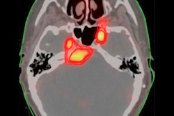
 Axial imaging of glomus jugulare and treatment planning. A) T1-weighted MRI with contrast. B) Ga-68 DOTATATE PET/CT. C) MRI-PET fusion. D) Gamma knife radiation dose distribution. Image courtesy of Patrick Wojtylak.
“Ga-68 DOTATATE PET/CT more effectively distinguishes between recurrent or residual tumors and scar tissue following surgery, offering higher sensitivity and specificity than MRI,” Wojtylak said.
Ultimately, the study included a small number of patients, but suggests Ga-68 DOTATATE PET/CT holds promise as a reliable imaging biomarker for HNPGLs, particularly as the incidence of NETs has increased over the past few decades and that imaging is crucial in the management of these tumors, Wojtylak noted.
“Integrating Ga-68 DOTATATE PET/CT into the clinical management of HNPGLs can lead to more accurate diagnosis, better treatment planning, and improved patient outcomes,” he concluded.
Wojtylak is the radiology system manager for nuclear medicine at the Cleveland Medical Center. The study was one of several discussed during a session of oral abstract presentations by technologists.