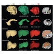
Combining deep learning with micro-CT shows promise for not only tracking the progression of lung disease but also evaluating the effectiveness of treatment, according to a study published May 9 in Respiratory Research.
Why? In part because deep learning automates the postprocessing phase of CT image analysis, noted a team led by Martina Buccardi, PhD, of the University of Parma in Italy.
"[Micro-CT] has proven to be a powerful tool to visualize and precisely quantify the dynamic evolution and regional severity of disease," the investigators wrote. "However, to take full advantage of this approach, it is necessary to automate the postprocessing phase of analysis, including lung segmentation."
CT is the gold standard for evaluating lung disorders, and its "miniaturized" version -- micro-CT -- has been shown to be an equally effective, noninvasive tool for tracking lung disease progression and monitoring the performance of new drugs for pulmonary disease in small animal studies. But it still requires manual image processing -- which can be not only time-consuming but susceptible to vagaries in operator skill, the group noted. That's where artificial intelligence (AI) and deep-learning algorithms come in.
"Manual segmentation ... has two main drawbacks that severely hinder its application to large datasets: It prolongs postprocessing time by up to 40 minutes per scan and it is prone to operator-dependent bias," the group noted. "These shortcomings have recently been addressed through the development of AI- and deep learning-based algorithms for automated lung segmentation in [animal] models of lung cancer and parenchymal pulmonary diseases."
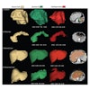
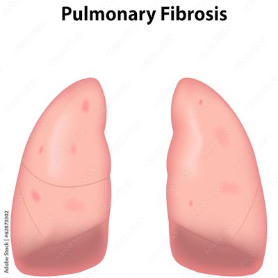
Via a mouse study of bleomycin-induced lung fibrosis, the investigators explored whether adding deep learning to micro-CT imaging in would improve and speed lung segmentation and the measurement of morphological and functional biomarkers in the whole lung and in lung nodes. (Bleomycin is a chemotherapy drug that can cause lung diseases.) The study included 19 mice who were administered bleomycin; of these, 12 were selected to receive the fibrosis-treatment drug Nintedanib (which is approved for human use). All the mice underwent micro-CT; the researchers processed these images with a deep-learning segmentation algorithm (developed in 2022 by Elena Vincenzi, PhD, and colleagues).
The team found that the combination of deep learning and micro-CT produced results consistent with conventional analyses (i.e., histology), and cut by 45-fold the time needed to analyze the imaging dataset and follow the development of fibrosis and its response to Nintedanib.
The investigators noted three benefits to this combined approach:
- Reduced experimental variability: Each animal subject is its own control and "the measured, operator bias-free biomarkers can be quantitatively compared across experiments," the group wrote.
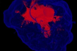
- Ability to monitor the spatial distribution of fibrotic lesions, thus "eliminating potential confounding effects associated with the more severe fibrosis observed in the apical region of the left lung and the compensatory effects taking place in the right lung," the authors noted.
- Potential to be used in drug discovery pipelines with "a substantial increase in both the speed and robustness of the evaluation of new candidate drugs," Buccardi's group explained.
The study findings show promise for the evaluation of new lung disease treatments, according to the researchers.
"The [micro-CT]-DL approach ... lends itself as a powerful new tool for the precision preclinical monitoring of bleomycin-induced lung fibrosis and other disease models as well," they concluded. "Its ease of operation and use of standard imaging instrumentation make it easily transferable to other laboratories and to other experimental settings, including clinical diagnostic applications."
