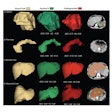
A brain imaging technique called magnetoencephalography (MEG) has identified a neural network that plays a key role in face pareidolia – a tendency to see faces in nonface images, according to neuroscientists in Germany.
The finding provides a framework for further research in patients with mental and neurological conditions, such as autism spectrum disorders, schizophrenia, and Parkinson’s disease, noted study lead Marina Pavlova, PhD, of Eberhard Karls University of Tübingen.
“Gaining knowledge about the specific patterns of possible alterations in brain communication in these patient populations will provide unique insights into the origins of social cognition deficits,” the group wrote. The study was published April 8 in Proceedings of the National Academy of Sciences.
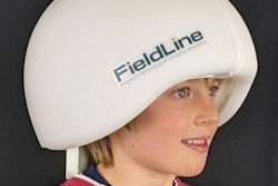
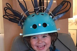
MEG is a noninvasive brain imaging technique that visualizes gamma oscillations, which are rhythmic patterns of neural activity, the authors explained. Gamma oscillations above 30 Hz are linked to a wide range of cognitive abilities, such as working memory and selective attention, and also underpin processing of social signals such as faces, they added.
The authors explored several questions, such as “What happens in the brain during face pareidolia?” and “Is processing of face-like images similar to that of faces?” They noted that to date, studies on the brain networks involved in face pareidolia are sparse and the outcomes have been controversial.
Thus, in this study, the group used a MEG unit (VSM MedTech, Coquitlam, BC, Canada) to study brain activity in 22 men on average 36 years old who were shown nonface face-like images and had to indicate the presence or absence of an impression of a face by pressing a key. None of the participants had a history of neurological or mental disorders.
According to the analysis, differences during processing of images that did or did not elicit face pareidolia occurred among the participants at later processing stages in the high-frequency gamma range of 80 to 85 Hz. Specifically, source localization analysis of the signals revealed multiple clusters of increased gamma activity in the right posterior superior temporal sulcus (STS-R), right inferior temporal gyrus (ITG-R), right insular gyrus (INS-R), right postcentral gyrus (PoG-R), and the left inferior parietal lobule (IPL-L). The greatest peak was found in the INS-R, the authors found.
 Examples of the Face-n-Thing images with canonical upright (top) and inverted (bottom) display orientation. The image on the left is one of the least resembling a face with upright display orientation, and the image on the right is one of the most resembling a face when presented with upright orientation. Image courtesy of Proceedings of the National Academy of Sciences.
“The key areas of the social brain, in particular, the STS and INS in both brain hemispheres, play a decisive role (like two kingpins) being strongly engaged in communication with other brain regions,” the researchers wrote.
Ultimately, the identification of communication signals involving the STS and INS is significant, as it suggests a framework for further investigations. The STS is likely the “top dog” in the face pareidolia network, and the INS is likely associated with the emotional aspects of face processing that habitually accompany the condition, the group wrote.
“The outcome provides a framework for future brain imaging research in patients with mental and neurological conditions characterized by aberrant face pareidolia (and face processing at large),” the group concluded.
The full study is available here.


