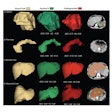
What are the most common clinical applications for 3D-printed models created from MRI scans of pediatric patients? A team from Ohio explored the various applications and the role radiologists play in production of the models in a new article, recently published online in the Journal of Magnetic Resonance Imaging.
"We wanted to investigate 3D printing with MRI because most applications using the technology have used CT scans in the past," first author Jayanthi Parthasarathy, PhD, told AuntMinnie.com. Parthasarathy is manager of 3D printing at Nationwide Children's Hospital's department of radiology in Columbus, OH.
"In children, you also want to avoid radiation as much as possible," she said. "So our goal was to see what were the most common applications in the pediatric world for 3D printing with MRI, and if it could give us the same benefits as 3D printing with CT does."
To that end, the group surveyed all 3D printing articles that were listed in the PubMed database from 1990 to 2019. They found 92 publications focused on pediatric 3D printing based on MRI scans as of March 2019. The vast majority of the articles were published in the last five years, with a peak of 19 articles published in 2017 (J Magn Reson Imaging, July 22, 2019).
The researchers for these prior studies most frequently used 3D-printed models for cardiovascular applications (35%), followed by use in neurosurgery (14%), education and training (13%), imaging (8%), and urology (8%), among others. Parthasarathy and colleagues discussed the specifics of several of these applications in their review:
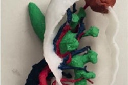
- Cardiology: 3D-printed models based on MR angiography images primarily serve to improve clinicians' understanding of the unique and complex arrangement of patient vasculature, which has proved critical for presurgical planning, the authors noted. The enhanced, tangible visualization of young patients' hearts has proved especially valuable in the management of congenital heart disease and for simulating interventional cardiology procedures.
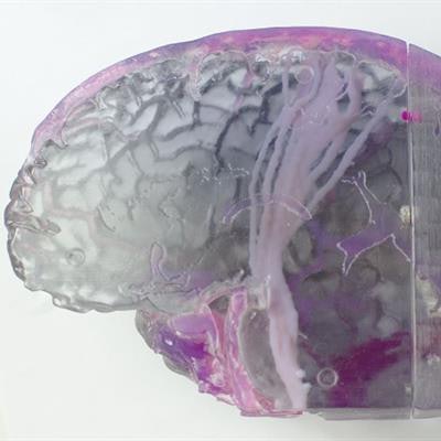
- Neurosurgery: Neurologists and neurosurgeons have used 3D-printed brain models as an adjunct during surgical planning for sensitive conditions such as intractable epilepsy. The models can display the entirety of the nerve tract in the brain (fusing multiple MRI contrasts in a single print), which has allowed for realistic, cost-effective simulation of challenging surgeries, according to the researchers.
- Education and training: Beyond surgical planning, 3D-printed models have also helped medical staff and patients and their family members come to understand anatomy more readily than by examining conventional medical imaging and even virtual 3D models.
- Imaging: Various groups have additionally used 3D printing technology to determine the best imaging sequences for MRI protocols.
- Urology: A number of investigators have demonstrated 3D-printed urologic cancer models to facilitate preoperative planning as well as improve patients' understanding of their condition.
The wide range of MRI-based 3D printing applications for pediatric cases has largely depended on collaborations between hospitals and external 3D printing services that necessitate long processing times, Parthasarathy said. However, point-of-care manufacturing for 3D-printed anatomical models, often led by radiology departments, has become increasingly common in recent years.
Better imaging techniques, demonstrated benefits of the enhanced visualization provided to surgeons and patients before surgery, availability of low-cost printers, and new 3D printing materials that better resemble human tissue have all contributed to this trend, and "new reimbursement pathways will add to a multifold increase in the utilization of the technology," she said.
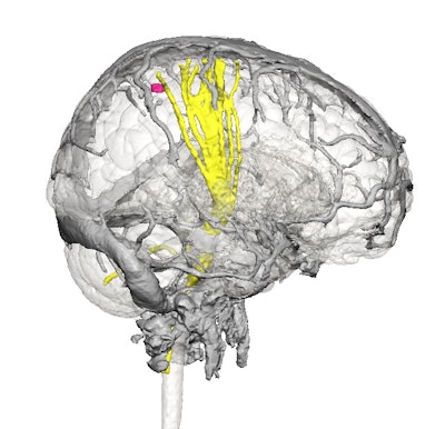 A virtual 3D model based on brain MR images (above). A patient-specific 3D-printed brain (below). All images courtesy of Jayanthi Parthasarathy, PhD.
"Radiologists are the people who do all of the imaging, and, as of now, the 3D printing applications at most hospitals are started in the radiology department," Parthasarathy said. "In a sense, the radiologist is the person who is the brains behind all of this. And in fact, radiology has taken up 3D printing in a big way."


