Contrast-enhanced mammography (CEM) has tradeoffs when it comes to imaging women with extremely dense breasts, according to research published October 1 in Radiology.
A team led by Maxine Jochelson, MD, from the Memorial Sloan Kettering Cancer Center in New York found that CEM demonstrated high sensitivity but lower specificity when compared with standard mammography images. However, CEM’s specificity increased on follow-up exams.
“The increased specificity of follow-up CEM examinations compared with baseline CEM examinations suggests that the diagnostic performance of CEM may be improved even further in the future,” Jochelson and colleagues wrote.
Standard mammography is limited in its ability to detect cancers in women with extremely dense breasts. This problem has led to researchers exploring different methods for better accuracy in imaging these women. CEM is one such modality that has gained attention in recent years, with the contrast enhancement highlighting suspicious findings better.
However, Jochelson and colleagues noted that data is limited on CEM’s use in women with extremely dense breasts. The researchers evaluated CEM’s diagnostic performance in this population, collecting data from consecutive CEM exams in asymptomatic women performed between 2012 and 2022.
From the CEM exams, the team defined "low-energy" images as the equivalent of a 2D full-field digital mammogram. It also used a postprocessing algorithm to recombine images highlighting areas of contrast enhancement and then compared results from both CEM and low-energy images.
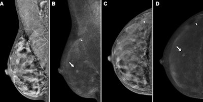
 Contrast-enhanced mammography (CEM) images depict a 56-year-old woman with a history of atypical ductal hyperplasia show cancer. Images include the following: screening (A, B) mediolateral oblique views and (C, D) craniocaudal views on (A, C) low-energy images and on (B, D) recombined images in the right breast. At the central slightly outer area in the right breast, mid-depth, there is a new enhancing mass (arrows) with no correlate on low-energy images. Subsequent targeted ultrasound showed a 0.5-cm irregular hypoechoic mass (not shown) for which ultrasound-guided biopsy showed invasive carcinoma, with a 1.2-mm invasive component at surgical pathologic examination and with negative nodes. Image courtesy of the RSNA.
The study included 1,299 screening CEM exams from 609 women with an average age of 50 years. The exams yielded 16 screen-detected cancers, with two interval cancers occurring. Of the cancers found, CEM depicted 11 of them while standard mammography found the remaining five.
The researchers noted that CEM led to an incremental cancer detection rate of 8.7 cancers per 1,000 exams. Baseline CEM exams achieved a higher sensitivity, but lower specificity than that of standard mammography.
| Comparison between CEM, low-energy imaging in extremely dense breasts | |||
|---|---|---|---|
| Measure | Low-energy imaging | CEM | p-value |
| Sensitivity | 27.8% | 88.9% | 0.003 |
| Specificity | 96.2% | 88.9% | < 0.001 |
However, CEM’s specificity at follow-up improved to 90.7%. The study authors suggested that this may be because of the availability of previous images at follow-up rounds, to which imaging readers can compare contrast enhancement in baseline and follow-up images.
“Furthermore, as clinical experience with CEM increases, it may be possible to avoid additional evaluation and/or biopsies for non-enhancing masses and asymmetries,” they wrote.
The study authors highlighted that while large-scale validation studies are needed, their data “shows the potential of CEM to address the unmet diagnostic challenge of breast cancer screening in women with extremely dense breasts with potentially more cost-effectiveness and availability than MRI.”
Marc Lobbes, MD, PhD, from the Zuyderland Medical Center in Geleen, the Netherlands, wrote in an accompanying editorial that while CEM continues to gain ground in radiological research, conducting large randomized controlled trials for this modality remains a challenge.
“Luckily, progress is already being made in that field, with several large prospective trials currently underway,” Lobbes wrote. “These large prospective trials will certainly help us identify who will benefit the most from screening CEM.”
The full study can be found here.


