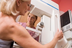
Combining full-field digital mammography with ultrasound tomography can improve breast cancer detection and be helpful in imaging dense breasts, according to research published June 18 in Radiology.
A team led by Mary Yamashita, MD, from the University of Southern California in Los Angeles, found that the combined method outperformed full-field digital mammography by itself, including having higher sensitivity for BI-RADS 4 lesions and higher specificity for BI-RADS 3 lesions.
“Our study suggests ultrasound tomography can be another supplemental screening tool,” Yamashita and colleagues wrote.
Previous studies suggest that women with dense breasts benefit from supplemental ultrasound imaging. However, ultrasound has low specificity and automated breast ultrasound (ABUS) prolongs interpretation time.
However, ultrasound tomography provides four coronal volumetric image sequences of B mode and quantitative tissue characterization information. This includes tissue stiffness, speed of sound, and attenuation.
“By merging reflection images with these parameters, ultrasound tomography can potentially differentiate cancer from normal tissue or benign disease in women with dense breasts,” the researchers wrote.
Yamashita and colleagues evaluated the performance of ultrasound tomography combined with full-field digital mammography and compared it with that of full-field digital mammography alone for screening women with dense breasts.
The study included data collected between 2017 and 2019 from 140 women who underwent both modalities at 10 centers as part of a prospective case collection registry. For the study, 32 radiologists who are qualified under the Mammography Quality Standards Act independently evaluated full-field digital mammograms followed immediately by mammography plus ultrasound tomography for suspicious findings and assigned a BI-RADS category.
Of the 140 women (average age, 56 years), 36 had breast cancer and 104 did not.
The team reported that the combined approach demonstrated superior performance compared with mammography alone. Additionally, combining mammography with ultrasound tomography led to superior sensitivity in BI-RADS 4 lesions and superior specificity in BI-RADS 3 lesions.
| Performance of full-field digital mammography plus ultrasound tomography, full-field digital mammography alone | |||
|---|---|---|---|
| Measure |
Mammography alone |
Combined approach |
p-value |
| AUC |
0.54 |
0.6 |
0.03 |
| Sensitivity (BI-RADS 4) |
30% |
37% | 0.03 |
| Specificity (BI-RADS 4) |
88% |
82% |
0.004 |
| Sensitivity (BI-RADS 3) |
33% |
40% |
0.08 |
| Specificity (BI-RADS 3) |
69% |
75% |
0.04 |
The radiologists performed 4,480 readings using both the combined approach and mammography alone. Of the readings, 4,475 combined approach readings and 4,332 mammography-alone readings took less than 30 minutes to complete.
The team reported that among images read in 30 minutes or less, the median reading time was 3.4 minutes for mammography plus ultrasound tomography and 1.2 minutes for mammography alone. However, for mammography plus ultrasound tomography, images with normal findings had a median reading time of 3.2 minutes compared to 3.9 minutes for mammography alone.
 Images from the automated breast ultrasound tomography system (SoftVue, Delphinus Medical Technologies) show four coronal volumetric image-stack sequences: wafer (waveform-enhanced reflection, which enhances fat so that dark masses are better visualized), sound speed (a direct output of image acquisition showing the change in the speed of sound moving through breast tissue), reflection (equivalent to B mode), and stiffness fusion (transmission properties of sound speed and attenuation overlaid on coregistered reflection images to highlight relative differences in tissue stiffness). A single slice from each sequence is shown.RSNA
The study authors wrote that while their results indicate ultrasound tomography’s utility in this area, more studies are needed to evaluate the modality’s benefits. This includes determining whether ultrasound tomography can improve cancer detection without increasing the number of benign biopsies.
In an accompanying editorial, Ritse Mann, MD, PhD, from Radboud University Medical Center in Nijmegen, the Netherlands, wrote that the results demonstrate ultrasound tomography being “at best on par” with handheld ultrasound and automated breast ultrasound. This includes allowing detection of some additional cancers at the price of a substantial increase in the number of false-positive findings.
“It seems that ultrasound tomography may be a new implementation of ultrasound, but it unfortunately yields old results,” Mann wrote.
Mann added that it remains to be seen whether “the promise of ultrasound can ever be delivered and whether ultrasound tomography has a role in that.”
The full results can be found here.