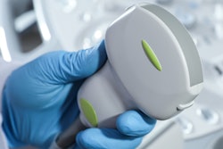
Ultrasound can help zero in on changes in back muscles that could be a sign of low back pain in healthcare workers who have to manually move patients, according to a study published February 11 in the International Journal of Industrial Ergonomics.
A team led by Robert Larson from Texas Tech University used ultrasound and electromyography to measure the size of the cross-sectional area of the multifidus muscle in the back. The group found that healthcare workers with smaller cross-sectional areas also reported higher pain scores.
"Because of the correlation between cross-sectional area and low back pain, increasing the multifidus cross-sectional area could be a way to reduce low back pain in this population, although the cause-and-effect relationship still needs to be explored," Larson and colleagues wrote.
Low back pain is a problem commonly seen in healthcare workers, a condition that can be exacerbated among those who have to physically move patients. Previous research suggests that some hospital workers can move up to 1.8 tons of patient weight during an eight-hour shift.
One of the most common tasks is boosting patients up and into a bed. The forces that go into this task have been studied when it comes to injury, but they have not been linked to pain experienced every day.
Previous research has also looked at how the cross-sectional area of the multifidus muscle plays into low back pain. The multifidus is a group of triangular-shaped, muscle bundles in the deep back that range from the neck to the lumbar region. Measurements of the multifidus cross-sectional area can signal deconditioning or hypotrophy of the muscle, pointing to abnormality.
Musculoskeletal ultrasound is typically used to image and measure cross-sectional areas of these muscles. Electromyography is also used to measure muscle activity.
Larson et al wanted to look at factors that lead to low back pain in healthcare workers. These include whether higher peak forces experienced by workers when boosting patients affects pain levels, how the size of the multifidus cross-sectional area in workers plays into pain they experience, and how patients exhibit pain with different muscle activation patterns in the lumbar multifidus region.
The team looked at data from 35 healthcare workers of varying occupations and experience. Workers were also split into two groups, one experiencing pain and one nonpain. The nonpain group (17) had a visual analog score (VAS) of 0, while the pain group (18) had a score between 1 and 5.
Ultrasound was used to image the sacral and lumbar multifidus areas with workers lying in a prone position. Workers also raised their legs upon instruction to engage the multifidus muscle for imaging.
The researchers found significant differences between the pain and nonpain groups when it came to ultrasound and electromyography-based measurements of the multifidus cross-sectional areas at the L5 and S1 spinal levels. Healthcare workers who marked lower pain scores on the visual analog scale had larger multifidus cross-sectional areas. The opposite was seen in workers with higher scores.
| Size of multifidus cross-sectional areas in healthcare workers experiencing pain | |||
| Spinal level | Pain group multifidus cross-sectional area (cm²) | Nonpain group multifidus cross-sectional area (cm²) | p-value |
| L4 | 9.88 | 10.6 | 0.0507 |
| L5 | 9.84 | 10.9 | 0.0360* |
| S1 | 8.49 | 10.2 | 0.0048* |
However, the team did not find any significant differences in peak low back force or multifidus activation between the pain and nonpain groups.
The study authors called for future research on the effects of strengthening the cross-sectional areas of the multifidus muscle in healthcare workers through exercise, as well as the use of electromyography sensors instead of surface sensors to measure pain.
"Comparing boosting techniques such as self-chosen bed height and posture between healthcare workers who have pain and those who do not have pain also has merit," they added.


