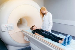
Patient sleepiness during a functional MRI (fMRI) scan distorts the brain connection maps produced by the exam, Mass General Brigham (MGB) researchers have found.
A team led by Cole Korponay, PhD, of McLean Hospital Imaging Center in Belmont, MA, (part of MGB) reported that "as people's arousal levels dwindle during an fMRI, such as if they become more relaxed and sleepy, changes in breathing and heart rates alter blood oxygen levels in the brain -- which are then falsely detected on the scan as neuronal." The group's findings were published June 19 in Nature Human Behaviour.
"You're lying down in a snug scanner for quite some time, often with only a low-engagement button press task to attend to or nothing to do at all, as the scanner monotonously hums and vibrates around you," Karponay said in a statement released by MGB. "These arousal-dampening conditions create the illusion that people's brain connection strengths continuously inflate throughout the scan."
Functional MRI measures neuronal activity by tracking changes in blood oxygen, making it vulnerable to "noise" from processes that affect blood oxygen such as breathing and heart rate changes, the group explained.
"And because breathing and heart rate patterns are closely tied to arousal levels, changes in arousal can introduce significant noise into fMRI data," it noted. "Problematically, the conditions of an fMRI scan tend to progressively lull people into lower arousal states."
Karponay and colleagues identified a specific blood flow signal dubbed the "systemic low-frequency oscillation" (sLFO) signal that indicated both the decrease in patients' arousal levels during the fMRI exam and the "illusory inflation of functional brain connection strengths." They noticed that the sLFO signal grew over time during the fMRI scan, in a spatial and temporal pattern that matched the pattern of the connection strength increases.
To correct for this, the team used a method called the Regressor Interpolation at Progressive Time Delays (RIPTiDe) method -- developed by co-senior study author Blaise Frederick, PhD, also at McLean -- to remove the signal from fMRI data and thus eliminate false brain connection strength increases.
The denoising procedure was effective for alleviating the "artifactual inflation of functional connectivity, enhancing the validity and within-scan reliability of fMRI findings," according to the authors.
"By adopting this sLFO denoising procedure, future studies can mitigate the distortive effects of arousal changes during brain scans and enhance the validity and reliability of fMRI findings," Korponay said in the statement.
The complete study can be found here.