Using 7-tesla MRI, Dutch researchers have identified the three most influential intracranial atherosclerotic risk factors associated with intracranial vessel wall lesions that lead to ischemic stroke, according to a study published in the April issue of Radiology.
Diabetes, hypertension, and increasing age -- in that order -- correlated with a higher number of vessel wall lesions in patients diagnosed with an ischemic stroke or transient ischemic attack (TIA). Perhaps most interestingly, the findings contradicted a trio of previously reported risk factors and their contributions to intracranial atherosclerosis and stroke.
"Although smoking, dyslipidemia, and male sex have been reported as independent risk factors for intracranial atherosclerosis in lumenography-based studies, no statistically significant association between these risk factors and vessel wall lesion burden was observed in our study," wrote lead author Dr. Arjen Lindenholz and colleagues from the University Medical Center Utrecht.
Risk factors such as hypertension, diabetes, and smoking have been associated with intracranial atherosclerosis, which accounts for 5% to 23% of ischemic strokes in the white population. While CT and MR angiography or digital subtraction angiography have been used to determine a link between intracranial calcifications, stenoses, and ischemic stroke, the modalities are limited in the evaluation of vessel wall lesions, which occur at the most advanced stage of intracranial atherosclerosis. Therefore, 3- and 7-tesla MRI have become increasingly utilized to detect the anomalies more expeditiously.
The question remains, however, as to which cardiovascular risk factors are the greatest contributors to vessel wall narrowing. In this study, Lindenholz and colleagues explored age, hypertension, gender, high or low levels of fat and lipids in the blood, and other influencers and their relationships to vessel wall lesions (Radiology, April 2020, Vol. 295:1, pp. 162-170).
Their analysis included 90 patients (52 men [58%]; mean age, 60 years; range, 27-85 years) who presented with ischemic stroke, transient ischemic attack, or transient monocular blindness at the Dutch facility between December 2009 and September 2017. The cohort included 46 cases (51%) of hypertension and 44 subjects (49%) with hyperlipidemia. In addition, 28 patients were smokers and 29 people had a history of smoking.
In correlating 7-tesla MR images of lesions with the risk factors, the researchers discovered three conditions that contributed most significantly to atherosclerotic lesions and stroke with anterior circulation vessel wall lesions:
- Diabetes mellitus, with a relative risk (RR) of 1.67
- Hypertension, RR of 1.46
- Increasing age, relative risk of 1.02
When the researchers considered vessel wall lesions in both anterior and posterior circulation lesions, the following were the significant risk factors:
- Diabetes mellitus, RR of 1.78
- Hypertension, RR of 1.49
- Higher systolic blood pressure, RR of 1.15
- Increasing age, RR of 1.02
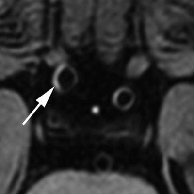
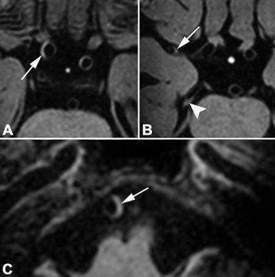
 MR images were acquired of a 69-year-old man with left-sided ischemic stroke 56 days after symptom onset. His cardiovascular risk factors were hypertension and diabetes mellitus. The Second Manifestations of Arterial Disease vascular risk score was 37%, indicating a 37% chance of a recurrent vascular event within 10 years. Fourteen vessel wall lesions were detected on pre- and postcontrast 7-tesla transverse 3D T1-weighted MRI. In the anterior circulation, postcontrast images (A, B) show vessel wall thickening (arrow in A) of the supraclinoid portion of the right internal carotid artery and right M1 segment (arrow in B). In the posterior circulation, postcontrast images (B, C) show vessel wall lesions in right P2 segment (arrowhead in B) and basilar artery (arrow in C). Images courtesy of Radiology.
In both analyses, the researchers found no statistically significant association among smoking (RR, 0.99), gender (RR, 0.84), and having abnormal lipid levels (dyslipidemia) (RR, 0.99) and the number of vessel wall lesions.
The lack of significant association might represent "vessel wall thickness fluctuation due to aging, natural variation, or partial volume effects, thereby resulting in underestimation of any association between the risk factors and 'true' atherosclerotic lesions," the study authors wrote. In addition, certain risk factors might only be associated with intracranial atherosclerosis "in specific ethnic populations or with more advanced stages of intracranial atherosclerosis, at which point luminal narrowing begins to develop."


