A pilot study conducted by German researchers and published in the August issue of Radiology suggests that PET/MRI provides diagnostic-quality images for assessing pulmonary masses in lung cancer cases, while cutting radiation dose by 75%.
In what is another boost for the prowess of the hybrid imaging modality, the study from Eberhard-Karls University in Tübingen also found similar lesion characterization and tumor staging in seven of 10 patients in a comparison of PET/CT and PET/MRI.
PET/MRI could provide another option for thoracic imaging, along with a reduction in radiation exposure from approximately 28 mSv for standard-dose contrast PET/CT to 7 mSv for whole-body PET/MRI, wrote lead author Dr. Nina Schwenzer, from the university's department of radiology, colleagues (Radiology, Vol. 264:2, pp. 551-558).
Improving upon PET/CT
The development of PET/CT has led to major improvements in the diagnosis of lung cancer, particular in the staging of both nodal and distant sites, the authors wrote. But the hybrid modality does have drawbacks, and MRI offers major advantages, such as better ability to visualize chest wall infiltration.
The arrival of hybrid PET/MRI scanners in the past year or so has raised the possibility of a simultaneous scan that could overcome the limitations of both PET/CT and MRI, they wrote. The team decided to compare PET/MRI's performance for lung cancer staging to that of PET/CT, and also to examine the quantification accuracy of the new modality.
Related story: MRI motion correction improves PET/MR image quality
The researchers enrolled 10 consecutive patients, ranging in age from 38 to 73 years, with a mean age of 59 years. The six men and four women were "highly suspected" of having or known to have lung cancer.
All 10 patients fasted overnight for at least 12 hours before receiving an intravenous injection of FDG. They were then given a whole-body contrast-enhanced PET/CT scan (Hi-Rez Biograph 16, Siemens Healthcare) from the skull base to the middle of the thigh.
PET/CT image acquisition began with contrast-enhanced CT after administration of 100 to 120 mL of iopromide (Ultravist, Bayer HealthCare Pharmaceuticals). PET imaging followed the CT scan 55 to 65 minutes after FDG injection with three minutes per bed position.
All PET/MRI exams were conducted using a whole-body hybrid PET/MRI system (Biograph mMR, Siemens) that can perform simultaneous PET and MR imaging during one examination.
The MRI portion of the device is a 3-tesla magnet with a length of 163 cm and a bore of 60 cm. The PET detector insert features lutetium oxyorthosilicate scintillation crystals in combination with MR-compatible photodiodes instead of photomultiplier tubes.
Related story: SNM: PET/MRI helps tumor staging for pediatric patients
Schwenzer and colleagues started PET and MR imaging simultaneously about 30 minutes after the PET/CT examination with six minutes per bed position. Uptake time after FDG injection ranged from 110 to 143 minutes, with a mean time of 126 minutes. PET image acquisition time per bed position was twice that of the PET/CT examination to make use of the MR image acquisition time.
The researchers also calculated tumor-to-liver ratios for all pulmonary tumors according to region of interest. For their volume-of-interest and region-of-interest analyses, PET datasets from PET/MRI and PET/CT were coregistered. Two readers then performed tumor staging based on PET/CT and PET/MRI results.
PET/MRI matches PET/CT
Examinations with both types of scanners were feasible in all 10 subjects, with PET/MRI producing "diagnostic image quality without apparent image artifacts" and "good tumor delineation," the authors wrote. The staging results also showed high agreement between PET/CT and PET/MRI in most cases.
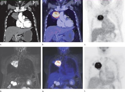 |
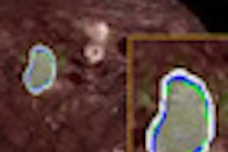
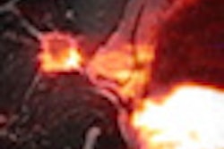
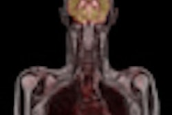
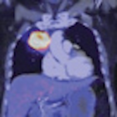
| Coronal PET/CT and PET/MR images of a 64-year-old woman with lung cancer in the right upper lobe. Anatomic images (a,d), superimposed PET and anatomic images (b, e), and FDG-PET images (c, f) illustrate the findings. Images courtesy of Radiology. |
In seven of 10 patients, PET/MRI provided an equivalent finding in the diagnostic assessment of pulmonary lesions when compared to PET/CT as reference. Nine of 10 lesions showed pronounced FDG uptake. One lesion was morphologically suspicious for malignancy at CT and MRI but showed no FDG uptake.
"Our preliminary data indicate that simultaneous PET/MR imaging offers an alternative modality in thoracic imaging with markedly reduced radiation exposure (approximately 75% reduction in dose) from about 28 mSv for standard-dose contrast material-enhanced PET/CT to 7 mSv for whole-body PET/MRI examination," Schwenzer and colleagues concluded.
They recommended further research into simultaneous PET/MRI in therapy monitoring of pulmonary masses, especially with regard to functional MRI techniques.


