A feasibility study in rats suggests that PET/CT can detect brain damage caused by trimethyltin chloride (TMT), a hazardous compound widely used in plastics production.
Researchers in Guangzhou, China, noted that kidney and cardiac toxicity from TMT poisoning is well established in humans, yet the study of neurotoxicity from the compound is still in its nascent stage.
“This study aims to provide a new evaluation method for diagnosing and treating TMT poisoning while shedding light on its underlying mechanisms,” wrote first author Anqing Liu, of the Guangdong Province Hospital for Occupational Disease Prevention and Treatment. The study was published January 8 in Scientific Reports.
TMT is a highly toxic chemical widely used as a heat stabilizer in industrial plastics production. It can cause kidney and liver damage, as well as mania, delirium, ataxia, epileptic seizures, and even death in poisoned humans. Previous studies using brain CT and MRI in exposed patients have reported low detection rates, however, and thus there is a need for a more sensitive brain imaging method, the authors explained.
To that end, the group explored the use of micro-PET/CT with F-18 FDG radiotracer to observe changes in overall brain glucose metabolism in rats exposed to TMT and normal control rats at different time points. They also studied neurobehavioral and neuropathological results, aiming to evaluate the feasibility of PET/CT in detecting and localizing nervous system damage.
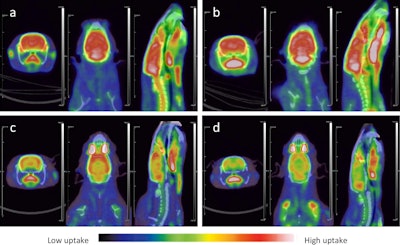
 Micro-PET/CT images of rats. (a) At 24 hours in the control group, F-18 FDG uptake in rats brain tissue was normal, and the PET/CT images were red and white; (b) At 7 days in the control group, F-18 FDG uptake in rats brain tissue were normal, and the PET/CT images were red and white; (c) At 24 hours after TMT exposure, F-18 FDG uptake in rats brain tissue were in a low state, and the PET/CT images were orange and yellow. (d) 7 days after TMT exposure, F-18 FDG uptake in rats brain tissue further decreased, and the PET/CT images appeared yellow and light yellow. There was no significant difference in the uptake of muscle tissue between the model group and the control group, and the PET/CT image was blue. Image available for republishing under Creative Commons license (CC BY 4.0 DEED, Attribution 4.0 International) and courtesy of Scientific Reports.
According to the results, TMT resulted in a significant reduction in F-18 FDG uptake across a broad range of brain tissue. F-18 FDG uptake was slightly reduced compared with the control group at 24 hours after TMT exposure, but by the seventh day, there was a significant decrease in F-18 FDG uptake in a wide range of brain regions. Further testing revealed that TMT caused pathological damage in the cerebral cortex, cerebellum, and hippocampus of the rats.
“PET/CT of TMT exposed rats had abnormalities in the early stage, and the abnormal brain functional areas were highly consistent with the symptoms of TMT neurotoxicity,” the group wrote.
Moreover, the exposed rats had spasms 30 minutes after TMT exposure, which disappeared several hours later, and starting from the second day after exposure, the rats had obvious aggression, irritability, and slight tremor of the head, the researchers added.
Ultimately, in humans, there is no obvious organ damage in the early stages of TMT poisoning, and PET/CT exams could serve to detect early abnormal functional changes in brain tissue in exposed patients, the group suggested. However, this was an animal study, and further research is warranted, they wrote.
“These results undeniably support the feasibility of utilizing PET/CT technology for detecting TMT poisoning, thereby suggesting its potential as a valuable tool for early diagnosis,” the researchers concluded.
The full study can be found here.


