PET/CT imaging with an F-18 DCFPyL prostate-specific membrane antigen (PSMA) radiopharmaceutical changed prostate cancer management in two-thirds of cases, according to research presented July 13 at the Society of Nuclear Medicine and Molecular Imaging (SNMMI) 2020 virtual annual meeting.
Previous research has suggested that PSMA-PET/CT is effective for diagnosing prostate cancer, but its impact on management of patients with suspected recurrent disease has been unclear, wrote a team led by Dr. Ur Metser of the University of Toronto in Canada. To address the question, the group conducted a study using PSMA-PET/CT that included 410 men with suspected recurrent cancer who had no disease on conventional CT or bone scintigraphy and who had undergone multiple prostate cancer treatments.
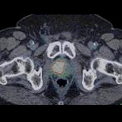
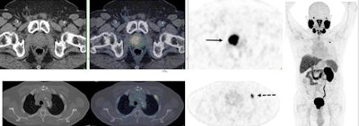
 PSMA-PET/CT accurately detects recurrent prostate cancer in 67-year-old man. F-18 DCFPyL-PSMA PET/CT shows extensive, intensely PSMA-avid local recurrence in prostate (bottom row; solid arrow) in keeping with known tumor recurrence in the prostate. Right: PET shows extensive, intensely PSMA-avid local recurrence in prostate (top row; solid arrow) and a solitary bone metastasis in left rib two (bottom row; dotted arrow). Image courtesy of Dr. Ur Metser of the University of Toronto; caption courtesy of the SNMMI.
Of the 410 men, 64% had PET-detected lesions. In all, 56% of men with negative conventional imaging results had lesions detected by PET; among those with lesions on conventional imaging, 63% were found to have new ones. PET/CT imaging also showed the following:
- 12% of men had localized disease.
- 23% had regional nodal without distant disease.
- 29% had distant disease.
- 35% had recurrent disease in the pelvis.
PSMA-PET/CT altered treatment in 66% of the men, the most common shifts being from observation or systemic therapy to surgery or radiation.
"The identification of extent of recurrence and specific sites of recurrence is crucial in determining the most appropriate mode of therapy for these men," Metser said in a statement released by the SNMMI. "Findings from this study add to the body of evidence on the utility of PSMA-PET in the management of prostate cancer patients."


