CAMBRIDGE, MA - Technology is affecting virtual colonoscopy in ways that will improve the patient experience and increase the technique's detection accuracy, according to presentations at the 2011 International Symposium on Virtual Colonoscopy.
Reviewing a few of the dozens of recent technology and research advancements presented at the symposium, Dr. Ronald Summers, PhD, director of radiology research at the U.S. National Institutes of Health (NIH), touched on innovations in virtual colonoscopy that could change practice patterns and differentiate VC from other colon screening tests.
His initial discussion excluded computer-aided polyp detection, a larger focus of technology research in virtual colonoscopy (also known as CT colonography or CTC) that was addressed in later presentations at the symposium.
Bone densitometry
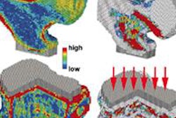
Bone mineral density measurements from CTC data continue to grow in sophistication and accuracy, and bone densitometry may prove to be a workable add-on test that increases the value of a virtual colonoscopy scan. The procedure is useful in roughly the same group of individuals 50 and older and at risk of colorectal pathology.
"Since the patient's already been scanned, you can get the bone mineral density without any additional radiation exposure," Summers said.
This year, Summers and his NIH team developed a fully automated technique to measure bone mineral density (BMD) that operates without the need for a calibration phantom (Journal of Computer Assisted Tomography, March-April 2011, Vol. 35:2, pp. 212-216). Fully automated BMD measurements were reproducible for a given patient with 95% limits of agreement of -9.79 to 8.46 mg/mL for the mean difference between paired assessments on supine and prone CTC, the group reported.
Dr. Perry Pickhardt, from the University of Wisconsin, and colleagues including Summers went beyond assessment to comparison with traditional dual-energy x-ray absorptiometry (DEXA) bone density measurements. The recent study compared their CT scores to a subset of patients undergoing DEXA scans, establishing both accuracy and a reference standard for the technique, Summers noted.
Sensitivity was 100% for osteoporosis, the authors wrote in the Journal of Bone and Mineral Research, though specificity was substantially lower at 46%.
"It's sensitive for picking up all the patients with osteoporosis, but it overcalls," said Summers of the system, which can be adjusted if needed to decrease detection sensitivity in exchange for boosting specificity. CT offers inherent advantages over DEXA in its ability to distinguish different regions of the vertebral bodies, Summers said.
Audience members suggested that following disease progression might be a worthwhile project for validating the numbers further, and specificity might be improved with the use of dual-energy CT.
Visceral fat
Visceral fat analysis began at NIH mainly for assessing patients on certain drug therapies -- particularly protease inhibitors for HIV patients -- that can lead to lipodystrophy, according to Summers. Pickhardt and colleagues also showed that the technique is very useful for assessing patients at risk for metabolic syndrome, Summers said.
"You can easily do fully automated visceral fat analysis at CTC without any additional radiation exposure, or any change to the imaging protocol," Summers said. It's superior to body mass index (BMI) assessment, which cannot distinguish between visceral and subcutaneous fat. In a presentation at the 2010 RSNA meeting, Sussman and colleagues reported that in a cohort of 719 men, patients in the highest quintile of visceral fat volume had twice the odds ratio of having adenomatous polyps compared to the thinnest quintile.
Still, a metabolic fat index will need to be developed and validated before it can be incorporated into a package of bundled diagnoses based on CTC data, suggested Dr. Michael Zalis from Massachusetts General Hospital.
Supine-prone data registration
Automated registration of prone and supine datasets has the potential to save a lot of time for radiologists interpreting CTC studies, and several investigators are working on it, Summers said.
Among them, doctoral student Holger Roth and colleagues from University College London developed a technique that deforms supine images to match prone CTC data from one endoluminal surface to the other (Medical Physics, June 2011, Vol. 38:6, pp. 3077-3089).
The cylindrical registration technique uses surface curvature information as a measure of similarity between surfaces to propel registration to compensate for the colorectal deformations that occur between prone and supine CTC data acquisitions.
In another approach to polyp location matching -- this one presented at the Computer Assisted Radiology and Surgery (CARS) meeting -- Summers, Kevin Chang, PhD, and colleagues achieved accurate polyp localization using a method based on a normalized distance calculation along the colon center line. Because of the stretching and distortion that occurs during conventional optical colonoscopy, the colon is about twice the length as it is during VC, complicating the matching of lesions detected in one modality versus the other.
Speaking of his group's study, Summers said the method "monitors more closely the mechanical properties of the colonoscope -- its torsion and its limitations on the curvature -- and tries to correct for those properties as the scope is inserted." Using the technique, a greater proportion of the polyps were located to within 5 cm of their position at CTC, he said.
Electronic stool cleansing
In terms of recruiting more patients to colon screening, few developments have the potential of electronic cleansing (EC) of tagged fecal materials, which would permit individuals who strongly oppose cathartic bowel cleansing to get screened. The number of screening-age individuals who oppose cleansing enough to skip screening is unknown, but it's probably substantial.
Several techniques have been developed to distinguish colonic mucosa from residual stool, all of which are imperfect and produce some artifact. But significant improvements have been made in those systems in the past couple of years. Reduced cleansing regimens have been developed to maximize the results of the new algorithms.
Among those researching this area are Neil Panjwani, Summers, and colleagues, who presented an automated method of subtracting tagged fecal matter at the 2011 Medical Image Computing and Computer Assisted Intervention (MICCAI) meeting in Toronto, Summers noted. The method proposed is locally adaptive and combines intensity, shape, and texture analysis with probabilistic optimization, the researchers wrote in an abstract accompanying the presentation. Depending on the specific bowel preparation used, automated detection of polyps using the group's computer-aided detection (CAD) system on cathartic-free data improved the true-positive detection rate significantly, from 70% to 85% at 5.75 false-positives per scan.
Presenting at the same Toronto meeting, Wenli Cai, PhD; Hiroyuki Yoshida, PhD; and colleagues from Massachusetts General Hospital described their technique for minimizing partial volume effect (PVE), a major cause of artifacts in electronic cleansing. Their technique relies on dual-energy index (DEI) -- specifically, the high DEI of air and tagged fecal materials -- to identify voxels at the boundary between air and tagged materials versus those of soft-tissues structures, which have a DEI value near zero, the authors wrote in an abstract.
A Massachusetts General Hospital pilot study of minimally cleansed CTC patients presented at last year's RSNA meeting yielded greater than 90% detection sensitivity for polyps 9 mm and larger. A much larger study of 600 patients undergoing minimally cleansed VC using the Mass. General electronic cleansing algorithms is in the works, according to co-investigator Dr. Michael Zalis.
Teniae coli
Teniae coli are three bands of longitudinal smooth muscle tissue on the surface of the colon. Because they run parallel along the colon wall, they represent important anatomical landmarks for both polyp location and prone and supine image registration.
The muscles are equally distributed on the colon wall and form a triple helix structure from the appendix to the sigmoid colon, NIH investigators wrote in an abstract describing their work.
At this year's SPIE Electronic Imaging meeting in San Francisco, Summers and colleagues presented a fully automated method of detecting teniae coli. Combined with the center-line method, the technique can be used for more precise longitudinal and circumferential polyp registration, he said of the study led by his colleague Zhuoshi Wei. The technique was used to detect the teniae coli using height maps from CTC image data.
The method begins with an unfolding of the 3D colonic surface using a parallel projection technique, followed by computation of a 2D height map of the unfolded colon, the authors explained. The teniae coli are detected on the height map and projected back to the 3D colon. Gabor filters are then used to extract haustral folds to eliminate distortions based on the location of the teniae coli at haustral folds. In experiments carried out in seven cases, the technique yielded promising result with an average normalized root mean square error (RMSE) of just 5.6% and a standard deviation of 4.79% of the circumference of the colon.
Results within 5% are not only close enough for government work, they are accurate enough to use for developing systems that can accurately match prone and supine datasets in CTC.
"The ability to register prone and supine datasets ... would make my life teaching people how to do this a lot easier," commented symposium course director Dr. Matthew Barish from Stony Brook University Medical Center, taking the opportunity to urge the vendor representatives in attendance to consider adding a prone-supine registration technique to their toolkits.
Significant time is spent learning -- and debating -- how to determine whether a feature detected on one dataset is the same as a different-looking feature on the other. "To incorporate some of this technology into the software for reading would be a huge win ... for radiologists to feel more confident," Barish said. Expert readers have learned how to do it accurately, but novice and even moderately experienced readers could be spared the struggle with reliable prone-supine matching, he said.


