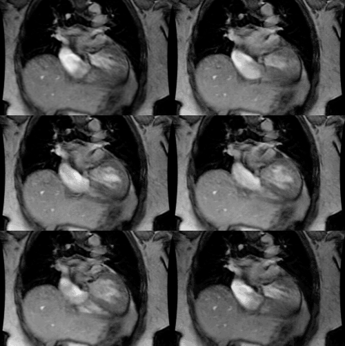
Hypoplastic Left Heart:
Clinical:
The term hypoplastic left heart (HPLH) describes a complex
collection of cardiac
anomalies involving hypoplasia or absence of the left ventricle.
In its most common (84%
of cases) and classic form there is aortic valve atresia, a
hypoplastic ascending aorta, a
hypoplastic/atretic mitral valve, and hypoplasia of the left
ventricle. In patients with
HPLH, returning pulmonary venous blood to the left atrium cannot
enter the left ventricle
due to an atretic or stenotic mitral valve. The blood is therefore
shunted to the right
atrium via an ASD where it is mixed with systemic venous return.
The interatrial defect is
most commonly related to a patent foramen ovale. In these cases,
blood cannot adequately
empty from the left atrium and this produces pulmonary venous
outflow obstruction. This
results in the typical presentation of the neonate with severe
congestive heart failure.
The right ventricle supplies blood to the pulmonary circulation.
Systemic arterial supply
occurs via a patent ductus arteriosus which is required for
survival- thus, the right
heart also supplies the systemic circulation. Unfortunately, the
right heart can neither
functionally or physiologically provide the volume of blood need
to sustain this type of
circulation [2]. The resulting systemic and coronary ischemia lead
to cyanosis and severe
acidosis [2]. Most patients present within the first few hours
after birth with CHF due to
pulmonary venous obstruction and RV overload. The presentation can
be delayed until 2 to 3
days of life if the ductus remains patent [1]. Hypoplastic left
heart is the most common
cause of CHF in the neonate. If untreated, almost all patients die
within the first month
of life.
Patients require a series of staged cardiac repair surgeries (Norwood procedure [3]) or cardiac transplant for treatment [1,2].
X-ray:
Prenatal ultrasound will demonstrate a small, hypoplastic left ventricle and diastolic flow reversal in an extremely narrow ascending aorta [2]. On CXR, there is usually enlargement of the cardiac silhouette, prominence to the right heart border, and findings of pulmonary venous hypertension/CHF [2]. Pulmonary arterial flow may be increased if the interatrial communication is large [2]. On angiography a string-like aortic coarctation is found in 80% of patients and there is retrograde flow from the RV via the PDA to the ascending aorta, arch, and coronary arteries. The right atrium is usually enlarged.
REFERENCES:
(1) Pediatric Clinics of North America 1999; Fedderly RT. Left ventricular outflow obstruction. 46 (2): 369-384
(2) Radiographics 2001; Bardo DM, et al. Hypoplastic left heart syndrome. 21: 705-717
(3) Radiology 2008; Gaca AM, et al. Repair of congenital heart disease: a primer-part 1. 247: 617-631


