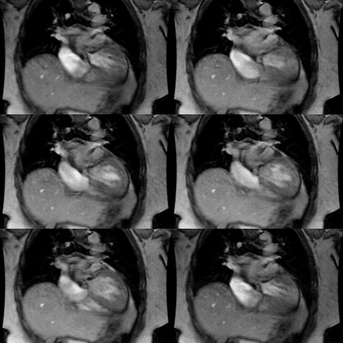
Patent Foramen Ovale
- Clinical:
A patent foramen ovale is a persistent valve-like connection between the left and right atrium [1]. During the first month of life this embryologic connection is usually closed via the formation of adhesions between the septum primum and secundum [1]. However, in about 25-27% of the population, the connection persists into adult life- resulting in a potential right-to-left shunt [1,2]. A left-to-right shunt is common in PFO, but any temporary elevation in right atrial pressure-such as with valsalva, cough, or strain- may provoke a right-to-left shunt [3]. In most people, there are no clinical consequences, however, PFO has been regarded as a potential source of cryptogenic stroke [4]. Also- in certain clinical instances- such as major pulmonary embolism or severe pulmonary hypertension, the shunt gains significance as an independent predictor of death or arterial thromboembolic (stroke) complications [1]. Patients with chronic obstructive pulmonary disease have an increased frequency of PFO because they have increased right atrial pressure compared to control subjects and the PFO can contribute to their chronic hypoxemia [2]. Atrial septal aneurysm is associated with PFO with a prevalence of 30-60%, and up to 64% of these patients can have a left-to-right shunt [3]. Transesophageal echocardiography (TEE) with agitated saline injection and Valsalva maneuver has previously been the gold standard for diagnosing PFO [2,4].
- X-ray:
MDCT provides detailed anatomic information regarding the size, morphologic features, and shunt grade of PFO [3]. A PFO produces a channel-like appearance to the interatrial septum, but only the presence of a contrast jet from the LA to RA confirms a PFO [4]. A shorter tunnel length (flap valve less than 6mm) and septal aneurysms are frequently associated with left-to-right shunts [3]. Some people have only a "flap valve" that appears as a thin line in the region of the foramen ovale- but if fused to the margin of the fossa ovalis, there is no opening that can produce a shunt [3].
REFERENCES:
(1) AJR 2008; Williamson EE, et al. ECG-gated cardiac CT angiography using 64-MDCT for detection of patent foramen ovale. 190: 929-933
(2) Radiology 2008; Revel MP, et al. Patent foramen ovale: detection with nongated multidetector CT. 249: 338-345
(3) Radiology 2008; Saremi F, et al. Imaging of patent foramen ovale with 64-section multidetector CT. 249: 483-492
(4) Radiology 2009; Kim YJ, et al. Patient foramen ovale: diagnosis with multidetector CT- comparison with transesophageal echocardiography. 250: 61-67


