Radiographics 1997 May;17(3):595-608. MR imaging of congenital anomalies of the thoracic veins.
White CS, Baffa JM, Haney PJ, Pace ME, Campbell AB
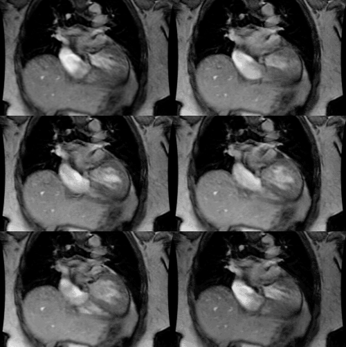
Congenital anomalies of the thoracic veins are infrequent but important developmental abnormalities. Thoracic venous anomalies can be classified as systemic or pulmonary. Systemic venous anomalies are often incidental findings, whereas pulmonary venous anomalies are more likely to manifest with cyanosis and to be associated with congenital cardiac abnormalities, especially atrial septal defect. Magnetic resonance (MR) imaging provides excellent delineation of the abnormal vessels and associated cardiac defects. Conventional spin-echo (SE) techniques show blood flow as a signal void and are sufficient for demonstrating the aberrant venous anatomy in most cases. Gradient-echo images show flowing blood as high signal intensity and are useful for clarifying the course of anomalous veins when vessel walls are difficult to visualize on SE images. Phase-contrast images are valuable for ascertaining the direction of blood flow and thus provide a physiologic method of distinguishing the vertical vein of anomalous pulmonary venous return from a left superior vena cava. MR imaging is useful for delineating both the thoracic venous and accompanying intracardiac anomalies and is a valuable, complementary technique to echocardiography, angiography, and computed tomography in the evaluation of patients with these abnormalities.
PMID: 9153699, MUID: 97298251


