AJR Am J Roentgenol 1996 Jun;166(6):1379-1385. The hepatopulmonary syndrome: radiologic findings in 10 patients.
McAdams HP, Erasmus J, Crockett R, Mitchell J, Godwin JD, McDermott VG
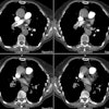
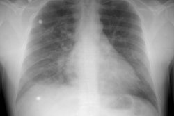
OBJECTIVE: The purpose of this study was to review the radiologic manifestations of the hepatopulmonary syndrome. MATERIALS AND METHODS: We retrospectively reviewed clinical records, chest radiographs, 99m Tc-macroaggregated albumin (MAA) perfusion lung scans, chest CT scans, and pulmonary angiograms of 10 patients with proven hepatopulmonary syndrome. RESULTS: Chest radiographs showed basilar, medium-sized (1.5-3.0 mm) nodular or reticulonodular opacities in all cases. CT was done in eight cases and showed basilar dilatation of lung vessels with a larger than normal number of visible branches. The vascular basis for these opacities was best appreciated on conventional CT scans of 10-mm sections. No individual arteriovenous malformations were seen on CT scans. High-resolution CT scans showed no evidence of interstitial fibrosis. 99mTc-MAA perfusion lung imaging, done in seven patients, showed pulmonary arteriovenous shunting in five. Contrast echocardiography confirmed intrapulmonary shunting in these five patients. Pulmonary angiography, done in four cases, showed subtle distal vascular dilatation in two and moderate dilatation with early venous filling in two but did not reveal any individual arteriovenous malformations. CONCLUSION: Chest radiographs in hepatopulmonary syndrome usually show bibasilar nodular or reticulonodular opacities. Conventional CT shows that these opacities represent dilated lung vessels. High-resolution CT is useful in excluding pulmonary fibrosis or emphysema as the cause of these opacities. 99mTc-MMA perfusion imaging or contrast echocardiography can be used to confirm intrapulmonary arteriovenous shunting.
PMID: 8633451, MUID: 96222705


