MRI can assess the entire ankle joint and exclude coexisting pathology in cases of posterior tibiotalar ligament (PTTL) injuries. In addition, ultrasound plays a complementary role in confirming instances of posteriomedial impingement that aren't immediately apparent on clinical exam, according to radiologists from Cabrini Private Hospital in Malvern, Australia.
"Severe ankle injury alone or in combination with inadequate treatment and/or rehabilitation can lead to impingement (which can be) an important cause of posteriomedial pain following ankle injuries...imaging plays a crucial role in early diagnosis and improving prognosis," wrote Dr. David Connell and colleagues in Foot & Ankle International (August 2003, Vol. 24:8, pp. 575-583).
For this study, conducted from October 1998 to August 2001, 25 consecutive patients with posteriomedial ankle pain were enrolled. The majority of injuries (14 to the right ankle; 11 to the left) were sustained while playing sports, including football and basketball; in five cases, the patients either fell down stairs or were involved in auto accidents. All of the patients complained of anteriolateral ankle pain, swelling, and inability to bear weight, the authors reported.
MR imaging was done on a 1.5-tesla unit (Signa LX, GE Medical Systems, Waukesha, WI). The ankle was immobilized with a surface coil, the toes upright and perpendicular to the long axis of the body. Axial, sagittal, and coronal fast spin-echo (FSE) imaging of the hindfoot was performed. The exam took 20 minutes.
Of the 25 patients, 17 also underwent 15-minute sonographic exams with either a 5-12 MHz transducer (HDI 5000, ATL, Philips Medical Systems, Bothell, WA) or a L10-12 transducer (Sequoia 512, Acuson, Siemens Medical Solutions, Malvern, PA).
  |
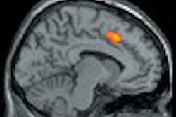
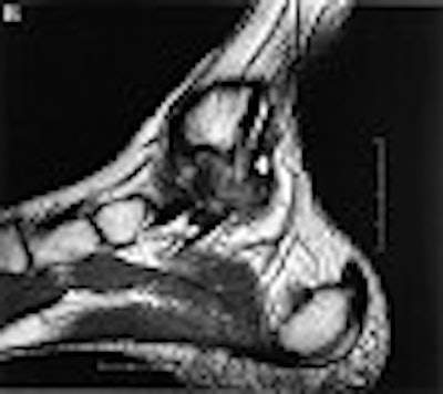
Coronal (A) and sagittal (B) MR images of PTTL injury
(arrow) with marked scar formation and hypertrophic response. The medial collateral ligament is lifted off the bone strictures of the medial ankle gutter by the volume scar formation. Encasement of the tibialis posterior tendon is noted (arrowhead). These findings are confirmed on ultrasound (C), which also detects the presence of calcific densities in the ankle gutter, either as a result of avulsion or dystrophic calcification.
Copyright © 2003 by the American Orthopaedic Foot and Ankle Society, Inc., originally published in Foot & Ankle International, 24(8): 575-583 and reproduced here with permission.
 |
The size of the deltoid ligament, and its relationships to the ligaments of the adjacent tendons, was evaluated based on morphology and abnormal MR signal, as well as morphology and echo texture on ultrasound. The surgeon had access to the imaging findings at the time of surgery.
According to the MR results, all 25 patients demonstrated a hyperintense posterior tibiotalar ligament, with the loss of a normal striated appearance and varying degrees of hypertrophy. On ultrasound, deltoid abnormality was found, which corresponded to the MR findings. Ultrasound also demonstrated a thickened hypoechoic ligament with diffuse loss of the normal fibrillar pattern, the group reported.
A dozen patients underwent surgery, at which time accessory loose scar tissue of degenerate posterior tibiotalar ligament was found, they added. The remaining patients were treated with a combination of conservative medication and corticosteroid injection at the site of impingement, explained co-author Dr. George Koulouris in an e-mail to AuntMinnie.com.
The authors stated that they found multiplanar MR imaging to be particularly helpful. Views of the ligaments in the coronal plane enabled accurate diagnosis of the posteriomedial impingement syndrome. They also found that ultrasound has value for demonstrating posteriomedial impingement lesions and small avulsion fractures.
"Following injury, a hypertrophic and inflammatory response can result in scar formation and inadequate healing," the authors wrote. "Identification of this lesion and exclusion of other causes of medial ankle pain can provide useful information to clinicians contemplating surgery."
Overall, the referring physicians expressed a preference for MR over ultrasound because the latter is more difficult to interpret, Koulouris said. At their institution, both modalities are not routinely performed for PTTL assessment; however they will do so at the request of the referring doctors. "Ultrasound does offer the advantage of correlation with site of direct tenderness as well as higher spatial resolution in a very small field," Koulouris added.
By Shalmali PalAuntMinnie.com staff writer
October 29, 2003
Related Reading
Just (over) Do It! Imaging pegs overuse injuries in specialty sports, October 8, 2003
MR categorizes acute hamstring injuries for pre-op assessment, September 29, 2003
The role of MRI and US in acute hamstring injuries: A preliminary report, July 22, 2002
Copyright © 2003 AuntMinnie.com