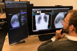
Who made it to the final round in this year's edition of the Minnies, AuntMinnie.com's event recognizing excellence in radiology? See below to find out who our expert panelists selected as the final candidates in this year's event.
The Minnies finalists were drawn from more than 200 candidates across 15 categories. You can also view a full list of the candidates, based on nominations from our members.
In the next round of voting, our expert panel will vote on the finalists, with winners announced in late October.
Most Influential Radiology Researcher
Maryellen Giger, PhD, University of Chicago
 Maryellen Giger, PhD.
Maryellen Giger, PhD, is arguably one of the best-known researchers in the field of computer-aided detection (CAD) and artificial intelligence (AI), so it's no surprise that the Minnies expert panel voted her as one of the two finalists in the Most Influential Radiology Researcher category.
Giger earned her Bachelor of Science degree at Illinois Benedictine College, received her master's degree in physics at the University of Exeter in England, and received her PhD in medical physics at the University of Chicago in 1985. Giger has been affiliated with the University of Chicago ever since, conducting some of the earliest studies in CAD applications in medical imaging.
At the university, she leads the Giger Lab, where she has advised more than 100 trainees over the years. Research programs underway at the lab include investigations into CAD and quantitative image analysis, digital radiography, MRI and MR spectroscopy, and x-ray tomography.
The rise of artificial intelligence in recent years has seen Giger's research take on even greater importance. In 2021, she was named to lead the Medical Imaging and Data Resource Center (MIDRC), a new initiative launched by the U.S. National Institutes of Health to spearhead the development of new diagnostic tools like AI algorithms to help diagnose and treat COVID-19. She discussed MIDRC in a talk at AuntMinnie.com's Spring 2021 Virtual Conference, Advances in AI.
Giger sees a major shift between the era of CAD and the emerging AI-based paradigm. For example, in its heyday, CAD was used mostly for breast screening in a second reader approach, while many of today's AI algorithms are designed to be used concurrently by radiologists as they interpret images. Still, she believes that today's AI developers can learn from the lessons of the CAD era, as she explained in a talk at the 2020 Conference on Machine Intelligence in Medical Imaging (C-MIMI).
In the past year, Giger's research has included papers on a machine learning algorithm to distinguish malignant from benign thyroid lesions on ultrasound, the creation of a "digital fetus" library for radiation dosimetry, and machine learning for early detection of ischemic brain injury after cardiac arrest.
Dr. Martin Pomper, PhD, Johns Hopkins University
 Dr. Martin Pomper, PhD.
Long known for his work in molecular imaging research, Dr. Martin Pomper, PhD, has appeared as a Minnies semifinalist candidate in the past, but this is the first year he has broken through to the finalist round -- perhaps an acknowledgment of his research productivity over the past year.
A professor in the neuroradiology division of the department of radiology at Johns Hopkins University, Pomper is also director of the Small Animal Imaging Resource Program (SAIRP) and co-director of the Johns Hopkins Center for Cancer Nanotechnology Excellence (CCNE). He is also co-director for PET at Johns Hopkins and is associate director of the In Vivo Cellular and Molecular Imaging Center (ICMIC) at the university. He serves as director of the university's Center for Translational Molecular Imaging (CTMI).
Pomper in particular has focused on the development of new molecular imaging radiotracers. He helped develop fluorine-18 (F-18) DCFPyL, a PET radiotracer that targets prostate-specific membrane antigen (PSMA) in cases of prostate cancer. The U.S. Food and Drug Administration approved the radiotracer in May 2021, and it is sold commercially as Pylarify by Lantheus Medical Imaging.
Over the past year, he has published a number of follow-up papers on F-18 DCFPyL, such as a publication in Journal of Nuclear Medicine in September 2022 that found that the agent changed patient management in nearly 50% of prostate cancer patients. Other papers Pomper has been involved with have discussed use of the agent in PET/CT to detect biochemical recurrence of prostate cancer after treatment, the best way to use the radiotracer to assess the success of therapy, and the best parameters to use when interpreting Pylarify images.
Pomper also conducted research into other radiopharmaceuticals, such as lutetium-177, which can also be used to target PSMA in prostate cancer cases. Further afield, he has also recently investigated near-infrared fluorescent imaging agents that target PSMA, as well as radiopharmaceuticals that target fibroblast activation protein alpha (FAP) in addition to PSMA.
Most Effective Radiology Educator
Dr. Christine "Cooky" Menias, Mayo Clinic Arizona
 Dr. Christine Menias.
It's not surprising that for the second year in a row, Dr. Christine "Cooky" Menias of Mayo Clinic Arizona has been nominated as Most Effective Radiology Educator. A specialist in abdominal imaging, body CT and MRI, and emergency radiology, she brings decades of experience to her educational efforts as professor of radiology at Mayo Clinic in Arizona. Last year, her educational activities further expanded when she began her tenure as editor of the RSNA's RadioGraphics journal.
Menias was born in Cairo, Egypt, and earned her medical degree at George Washington University School of Medicine and Health Services in Washington, DC. She has served as professor at the Mayo Clinic for almost a decade; before arriving at Mayo, she taught at Washington University School of Medicine in St. Louis for 17 years.
She has been often recognized for her educational leadership, snagging an Educator of the Year for Research award from the Mayo Clinic Arizona Resident and Fellows' Association in 2020; a Distinguished Educator award from the American Roentgen Ray Society in 2019; and the RSNA Honored Educator award, also in 2019 -- which she received each year beginning in 2014. In 2018, she received Mayo's Distinguished Clinician award, her "most cherished," since it highlights patient care and is rarely given to a radiologist, she said.
Dr. Stefan Tigges, Emory University School of Medicine
 Dr. Stefan Tigges.
At this point in his 30-year career at Emory University, Dr. Stefan Tigges has honed his skills in the radiology educator arena, and his teaching is wide-ranging and creative.
Not only is Tigges a professor of cardiothoracic imaging, he is director of the third year medical student radiology clerkship and an active teacher of medical students, residents, and fellows -- often using comics he draws and posts on his website, xraycomix or publishes in medical journals (topics range from the need for kindness in patient care to the vagaries of medical school training on Zoom).
In fact, last year he published a study in the Journal of the American College of Roentgenology that explored the benefits of teaching medical students about p-values via a graphic narrative compared with a journal article. When he teaches, he invites students to use drawing as they learn and stresses that it doesn't have to be complicated.
"Drawing is one of the most effective teaching methods available, especially if you have your learners draw along with you," he said in an article about his teaching published by the RSNA in 2021. "You don't need to make sophisticated drawings; it's been shown that novices learn better using simple drawings than more anatomically correct ones."
As for his own education, Tigges earned his medical degree from Emory, completed his residency at Vanderbilt University in Nashville, and returned to Emory for a musculoskeletal radiology fellowship. But it's his rather unorthodox approach to medical teaching that has gotten him noticed, most recently in 2020, when the Alliance of Medical Student Educators in Radiology bestowed on Tigges its Excellence in Education Award.
Most Effective Radiologic Sciences Educator
Kristi Moore, PhD, University of Mississippi Medical Center
 Kristi Moore, PhD.
When it comes to radiologic sciences education, Kristi Moore, PhD, sticks with a good thing when she finds it. Not only is she chair of the department of radiologic sciences at the University of Mississippi Medical Center’s School of Health Related Professions, but she also earned three degrees from the university, including a bachelor's in health sciences and both a master's and a doctorate in clinical health sciences.
As of late, Moore's educational activities have also included contributions to professional journals. Earlier this year, she coauthored a primer about how best to support students transitioning from general education courses to a specific radiologic sciences curriculum that was published in Radiologic Technology, and in 2021, she co-authored a study that explored the effect of MRI scanner noise on technologists, which was also published in that journal.
Currently, Moore is chair of the board of directors for the American Society of Radiologic Technologists (ASRT).
Louise Miller, Mammography Educators
 Louise Miller.
Louise Miller has lived out her deep commitment to radiologic sciences education -- specifically breast imaging -- through a panoply of initiatives and programs throughout her 35-year career. She has provided consulting to Harvard Medical School; the Mayo Clinic; the University of California, Los Angeles; and Stanford University -- as well as to more than 300 U.S. breast centers -- and has taught or trained an excess of 50,000 technologists across the globe. In 2013, Miller co-founded Mammography Educators, which offers mammography positioning training and education. In 1989 she co-founded and served as director of the Mammography Practicum at the University of California, San Diego.
Part of her educational efforts include three books: the Handbook of Mammography, 4th and 5th Edition in 1999; a positioning guidebook in 2014; and Image Quality & Positioning Problem Solving for Breast Imagers: Meeting EQUIP Standards in 2020.
Miller's dedication to radiologic sciences education has been acknowledged over the years. In 2011, she received the National Consortium of Breast Centers (NCBC) Impact Award, while in 2017, she was given the title of honorary fellow from the Society of Breast Imaging (SBI). Also that year, she was appointed consumer representative for the National Mammography Quality Assurance Advisory Committee, which counsels the U.S. Food and Drug Administration (FDA) on mammography-related quality issues and the enactment of the Mammography Quality Standards Act (MQSA).
Miller currently serves as a member of the American College of Radiology's (ACR) Mammography Positioning Improvement Collaborative.
Most Effective Radiology Administrator/Manager
Gina Greenwood, University of Wisconsin Hospital, Madison
 Gina Greenwood.
Gina Greenwood's skills as an administrator have been made plain throughout her 21-year stint at the radiology department at the University of Wisconsin in Madison, as evidenced by the variety of roles she has filled, from manager of MRI operations and clinical content facilitator to business operations manager, director of radiology, and now her current position as senior director of radiology.
Greenwood began her career as staff technologist at the University of Iowa in Iowa City and St. Luke's Hospital in Cedar Rapids, IA, and progressed to industry gigs with the likes of Lunar, GE Healthcare (then called GE Medical Systems), and IGC Medical Advances. While working on her bachelor's degree in health arts and a master's in business administration, she moved on to hone her administrative expertise at University of Wisconsin.
She's been committed to participating and supporting professional organizations in her field, which has also bolstered her administrative abilities. In fact, in a 2020 interview with the Association for Medical Imaging Management (AHRA) -- she was a candidate for director of the organization at the time -- she stressed the importance of maintaining strong networks, saying that her AHRA membership inspired her "to become more involved [and] make deeper connections."
Andrew Menard, JD, Johns Hopkins Health System, Baltimore
 Andrew Menard, JD.
As executive director of radiology strategy and innovation for Johns Hopkins Health System in Baltimore, Andrew Menard, JD, seeks to identify economic, political, and business trends that could affect both professional and technical radiology -- and then develop strategies to support the university's clinical care, education, and research.
Menard came to Hopkins in 2018 from Brigham and Women's Hospital in Boston, where he advised department chairs and vice chairs on policy, network and business development, and then served as executive director of radiology. While at Brighham he helped form a consortium to evaluate the impact of radiology clinical decision support on imaging utilization as part of the Medicare Imaging Demonstration, later providing input to draft legislation that eventually became the Protecting Access to Medicare Act (PAMA). He also assisted several academic radiology groups with their applications to become qualified provider-led entities under PAMA.
Menard's efforts have been noticed: In 2015, he received the Association of Administrators in Academic Radiology (AAARAD) award for advancement of the profession of radiology.