Ultrasound of the common extensor tendon produces high sensitivity, but low specificity for symptomatic lateral epicondylitis (tennis elbow), according to research from Thomas Jefferson University in Philadelphia.
To determine the sensitivity and specificity of ultrasound in this application, a TJU study team performed ultrasound of the common extensor tendon at the lateral epicondyle on 22 subjects with refractory symptoms of lateral epicondylitis. Fifteen patients received bilateral exams, three of whom had bilateral symptoms, said Dr. Dayna Levin in a presentation at the 2002 RSNA meeting in Chicago.
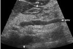
Bilateral ultrasound also was performed on 10 asymptomatic volunteers. All ultrasound studies were performed on a standard scanner with a high-frequency linear probe, she said.
A total of 57 hard copy images were generated from the studies, one from each elbow. Blinded to the clinical information, four independent radiologists rated each tendon as normal or abnormal.
For abnormal studies, the radiologists further classified the cases as demonstrating one or more:
- linear intrasubstance tears
- tendon thickening
- intratendinous calcifications
- adjacent bone irregularity
- focal hypoechoic regions
- enthesophytes at the tendon insertion site
- diffuse heterogeneity of the tendon
- peritoneous fluid
In addition, the same reader interpreted the same image on two different occasions to determine intra-rater variability, Levin said.
Ultrasound produced sensitivities of 92% (reader A), 84% (readers B and C), and 80% (reader D). However, specificity was 43.8% (reader A), 53.1% (reader B), 59.4% (reader C), and 40.6% (reader D).
"The low specificity may be explained by the fact that pathologic and sonographic changes occur before (the onset of) patient symptoms," she said.
The odds ratio (95% confidence interval) relating each ultrasound finding was statistically significant for each finding. Intratendinous tear, tendon thickening, and intratendinous calcification were the findings most associated with symptomatic epicondylitis, Levin said.
The odds ratio was 7.3 (1.5-34.6) for linear intrasubstance tears, 7.2 (3.8-13.5) for tendon thickening, and 6.1 (2.5-14.6) for intratendinous calcification. Adjacent bone irregularity had an odds ratio of 5.3 (2.2-12.4), while focal hypoechoic lesions had a ratio of 3.5 (1.7-7.3) and enthesophytes at the tendon insertion site had a ratio of 3.5 (1.7-7.1).
The odds ratio for diffuse heterogeneity was 3.2 (1.8-5.7), while peritendinous fluid was 2.4 (0.9-6.4). All odds ratios were statistically significant.
Both inter-rater and intra-rater correlation for overall impression was 0.38, Levin said.
"However, among two of the four readers with the most musculoskeletal ultrasound experience, inter-rater correlation was .63, which is very good," she said.
As for study limitations, Levin pointed out that their research studied a relatively small series of patients and that images were not interpreted in real-time. In addition, there was a lack of pathologic correlation. Finally, the sonographer’s skills represent a potential source of bias.
Because of the high false-positive rates, this exam will probably be most useful in patients with symptoms typical of lateral epicondylitis, rather than as a screening exam for patients with non-specific elbow pain, she said.
By Erik L. RidleyAuntMinnie.com staff writer
March 6, 2003
Related Reading
Boning up on humerus, clavicle, and AC joint positioning, February 18, 2003
US-guided needle therapy provides quick relief for joint injuries, December 5, 2002
The bends and flexures of forearm and elbow x-ray positioning, November 21, 2002
Ultrasound wins the gold at Sydney, November 8, 2000
Copyright © 2003 AuntMinnie.com