When it comes to meniscal tears of the knee, MRI can help radiologists home in on the exact source of pain. On the other hand, MRI may not be as valuable for traumatic knee injuries in an emergency setting. These are the conclusions reached by two studies presented at the 2002 RSNA meeting in Chicago.
First, investigators from Zurich, Switzerland conducted a prospective MR study of patients with clinically suspected meniscal tears.
"MRI is highly accurate for diagnosis of meniscal tears, with a reported sensitivity of 88% and a specificity of 94%. However, it’s also known that there is a baseline prevalence of meniscal tears in asymptomatic knees," said Dr. Marco Zanetti from the University Hospital Balgrist. The goal of the study was to determine which abnormalities are responsible for pain.
MRI was performed on the symptomatic knee, as well as the contralateral asymptomatic knee, in 100 patients with a mean age of 42.7 years (59 male; 41 female). Imaging was done on either a 1-tesla Magnetom Expert or a 1.5-tesla Magnetom Symphony scanner (Siemens Medical Solutions, Erlangen, Germany).
The imaging protocol included the use of "a dedicated circular-polarized, send-receive extremity coil on both MR scanners. The MR sequences were identical on both sides of each patient," Zanetti wrote in an e-mail to AuntMinnie.com. To learn more about the MR protocol used in this study, click here.
The prevalence of meniscal tears, abnormalities of the collateral ligaments, pericapsular soft tissues, bone marrow, and cartilage were determined. The meniscal tears were evaluated by two musculoskeletal radiologists and classified as follows:
- A - horizontal or oblique partial-thickness tears.
- B - radial tears.
- C - vertical or complex full-thickness tears.
- D - tears with dislocation of meniscal fragments.
According to the results, meniscal tears were found in 57 symptomatic knees and in 36 contralateral asymptomatic knees. In all 43 patients without a meniscal tear on the symptomatic side, normal menisci were found on the contralateral asymptomatic side. For those patients with a meniscal tear on the symptomatic side, the likelihood of having a tear on the contralateral asymptomatic side was 63%.
 |
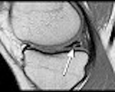
| The most common type of meniscal tear -- the horizontal or oblique partial-thickness tear -- is shown above (arrow). This type of lesion had a very similar prevalence on the asymptomatic contralateral side. |
Type A tears were found medially and laterally in 32 symptomatic knees and in 29 asymptomatic knees. Type B and C tears were seen medially and laterally in 18 symptomatic knees and in 5 asymptomatic knees.
Collateral ligament lesions were found in 52 symptomatic knees and in 7 asymptomatic ones, Zanetti reported. Pericapsular soft-tissue abnormalities were found in 63 symptomatic and 13 asymptomatic knees. Edema-like bone abnormalities appeared in 35 symptomatic and 4 asymptomatic knees. Cartilage lesions were present in 31 symptomatic and 25 asymptomatic knees.
 |
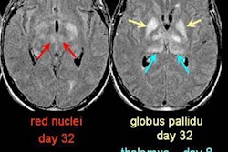
| Collateral ligament lesions (straight arrow), pericapsular edema (curved arrow) and edema-like bone marrow abnormalities (open arrow) were characteristic for the symptomatic side. Images courtesy of Dr. Marco Zanetti. |
The group concluded that type A tears are as common in the contralateral asymptomatic knee as in the symptomatic knee -- and that there is little evidence that such meniscal tears are responsible for pain. Instead, other types of meniscal tears, such as abnormalities of the collateral ligaments, pericapsular soft tissues, and bone marrow have a higher association with pain in the symptomatic knee.
While the group did not gather information on whether these MRI results influenced the treatment plan, Zanetti said that the feedback from orthopedic surgeons and rheumatologists has been positive.
"These results will prompt a more conservative treatment of meniscal tears," Zanetti said.
"We have planned to follow up these patients after two years to receive more information about the clinical relevance of meniscal lesions seen on MR imaging."
Dedicated MRI for knee trauma
In another presentation, Dr. Edwin Oei and colleagues from the Erasmus Medical Center in Rotterdam, the Netherlands, set out to determine the value of immediate dedicated extremity MRI after acute knee trauma. Specifically, they wanted to determine whether MRI results could predict the need for treatment within the follow-up period.
While MRI does have proven value for knee imaging, it can be cost-prohibitive, and is limited in availability in the Netherlands, Oei and co-authors wrote in their poster presentation.
The study population consisted of patients who visited the emergency department within seven days after sustaining a knee injury. They were randomized between radiographs only (the current workup at Oei’s institution), and x-ray plus immediate MRI. All imaging findings were reported immediately to the treating ER physician.
MR scans were performed on a 0.2-tesla dedicated extremity scanner (Artoscan M, Esaote Biomedica, Genoa, Italy). A short, five-pulse-sequence scanning protocol was used: spin-echo T1-weighted images were acquired in the sagittal-oblique plane. TME proton density-weighted plus T2-weighted images were acquired in the coronal plane; gradient-echo images also were acquired in the coronal plane; and STIR was used for imaging the sagittal plane. The total acquisition time was less than 15 minutes.
"We recorded if the patients had been treated during the follow-up period of six months after initial presentation. Univariable and multivariable logistic regression analyses were performed to evaluate patient characteristics, trauma mechanism, and findings on radiography and MRI," the authors wrote.
In total, 192 patients met the inclusion criteria from August 1999 to May 2001. Of the 192 patients, 111 required treatment such as arthroscopy or open surgery (27), immobilization (25), and physiotherapy (59).
MRI was predictive in the univariable analysis (odds ratios 0.44; 95% confidence interval) for a normal MR and for an abnormal MR (odds ratio 3.04; 95% confidence interval). However, the results for the multivariable models were not significant, the authors reported.
In comparison, abnormal x-ray was one of several significant predictors of treatment in both the univariable and multivariable analyses (odds ratio 4.91; 95% confidence interval), the authors wrote. Other influential factors were a patient age of 30 or higher (odds ratio 2.53: 95% confidence interval) and indirect trauma mechanism (odds ratio 4.42; 95% confidence interval).
The group concluded that MR might aid in predicting whether or not additional treatment is needed for a traumatic knee injury. However, it does not help identify patients who can be discharged without additional work-up or will not need follow-up treatment.
By Shalmali PalAuntMinnie.com staff writer
January 9, 2003
Related Reading
Turf Wars in Radiology, Part V: Radiologists, orthopedists put best foot forward, October 1, 2002
Dynamic MRI study differentiates leflunomide versus methotrexate for RA, March 14, 2002
Scenes from the Polyclinic: a case study, February 19, 2002
Olympic skiers tear up slopes -- and knees, February 18, 2002
Figure skating and stress fractures: A case study, February 14, 2002
Copyright © 2003 AuntMinnie.com