Magnetic resonance spectroscopy is more sensitive than MR imaging for gauging the lingering injury to newborns caused by hypoxic ischemic encephalopathy (HIE), according to an initial study from India.
The team from the Sanjay Gandhi Post-Graduate Institute of Medical Sciences in Lucknow published their findings in the European Journal of Radiology. The study compared MRI and MRS findings for 21 full-term neonates: 16 with hypoxic ischemic encephalopathy and five healthy controls.
HIE was diagnosed and graded by Sarnat stages based on clinical signs and a history of asphyxia at birth. Five of the neonates were deemed to be Sarnat stage I, with mild injury characterized by hyperalertness and hyperexcitability; nine were Sarnat stage II, with lethargy, hypotonia, and suppressed reflexes; and two were at Sarnat stage III, lacking normal movement and reflexes.
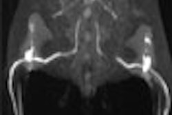
MR imaging and spectroscopy were performed in one session using a 1.5-tesla superconducting scanner (Siemens Medical Solutions, Erlangen, Germany) with a circularly polarized head coil. Conventional spin-echo T2- and T1-weighted imaging was performed in the axial plane using 3-mm slice thickness.
Single-voxel volume-selective spectroscopy was performed using an SE sequence with a voxel size of 8 ml. The researchers selected the voxel covering the area considered to be most sensitive to the effects of acute amoxia, the thalami, and basal ganglia. Both MRI and MRS findings were subsequently correlated with another clinical evaluation of the infants at two months of age, the authors wrote.
Although MR imaging is more sensitive than ultrasound or CT in detecting HIE-related abnormalities, the researchers stated, "the presence of normal imaging does not rule out neurodevelopmental delay," (EJR, July 2002, Vol.43:1, pp.6-13).
This point regarding the insensitivity of MRI in this area was borne out by the researchers’ own findings. In their study, MR imaging found no abnormalities in one of the Sarnat stage III neonates (both of whom had died by two months); conversely, two-thirds of the surviving neonates with MR imaging abnormalities had a normal clinical outcome at two months.
The few studies done previously on MRS in neonates with HIE have described wide-ranging but often inconsistent metabolic abnormalities in the brain, the authors noted.
"We believe that this discrepancy in these metabolite abnormalities among different studies is probably related to the location of the voxel selection, size of the voxel selection, and nonconsideration of the presence or absence of imaging abnormalities in the selected voxel," they said.
For their part, the Lucknow team found an increased concentration of intracerebral glutamate/glutamine (a -Glx) and glycine correlated with the severity of disease as determined by Sarnat staging. They also found that the presence of lactate was usually associated with a poor clinical outcome of death or neurodevelopmental sequelae.
"In the present study, the major advantage of MR spectroscopy in these neonates was that it allowed better delineation of intracranial abnormalities in the early stage of the disease even in the absence of imaging abnormalities," the authors concluded. "It shows that conventional T1- and T2-weighted MR imaging is relatively less sensitive than spectroscopy in predicting tissue ischemia in HIE."
By Tracie L. ThompsonAuntMinnie.com contributing writer
August 16, 2002
Copyright © 2002 AuntMinnie.com