Gallium-68 (Ga-68) DOTATATE PET/CT appears to be the best approach for detecting spinal bone metastases in patients with certain neuroendocrine tumors, according to researchers at the National Institutes of Health (NIH) in Bethesda, MD.
In a study of patients with pheochromocytomas and paragangliomas (PPGLs), the group found the approach was superior to F-18 FDG-PET/CT, MRI of the spine, whole-body CT, and whole-body MRI. The finding is significant regarding efforts to prevent serious complications in these patients, noted lead author Abhishek Jha, MD, and colleagues.
“Bone metastases weaken and destroy skeletal tissue and predispose cancer patients to acute and chronic skeletal-related events (SREs),” the group wrote. The study was published April 16 in European Radiology.
Bone metastases are frequently seen in patients with solid tumors and are observed in up to 71% of metastatic PPGL patients, the authors explained. SREs such as bone pain, spinal cord compression, and fractures not only affect morbidity and mortality rates in these individuals but also increase medical costs, they wrote.
However, data from head-to-head comparisons among imaging techniques to detect bone metastases in these patients are limited, the researchers noted.
To that end, the group enrolled 43 participants (mean age, 41.7) with diagnosed or suspected spinal bone metastases who had previously undergone MRI spine exams. All patients also subsequently underwent Ga-68 DOTATATE PET/CT (n = 43), F-18 FDG-PET/CT (n = 43), whole-body MRI (n = 24), and whole-body CT (n = 33) within six months.
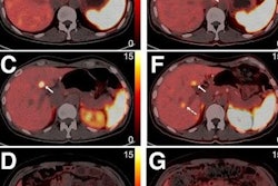
 Multimodality imaging of spinal bone metastases in a participant with pheochromocytoma and paraganglioma. The images of (a) whole-body Ga-68 DOTATATE anterior maximum intensity projection (MIP) and (b) fused sagittal PET/CT; F-18 FDG MIP (c) and fused sagittal PET/CT (d); T1-weighted sagittal MRI of the cervical (e), thoracic (f), and lumbar (g) spine, and contrast-enhanced CT (h sagittal) of a 25-year-old woman. This figure shows superiority of Ga-68 DOTATATE PET/CT in the detection of additional spinal bone metastases at C2, C5, C7, T1-2, T4-8, L2, and L5 compared to F-18 FDG-PET/CT in detecting spinal bone metastases at T9 (arrow on fused sagittal image and not appreciated on MIP image) and L4 (not appreciated on MIP and fused sagittal images), and MRI spine at C6, T10-12, and L1 (arrows), respectively. The whole-body CT was read negative for any spinal bone metastases. This participant did not undergo whole-body MRI. Image and caption courtesy of European Radiology.
Forty-one of 43 participants were positive for spinal bone metastases, with 382 lesions detected among the imaging approaches for the head-to-head analysis. The images were interpreted either by an expert nuclear medicine physician (PET/CT), radiologist (whole-body MRI and CT), or neuroradiologist (spine MRI).
According to the findings, Ga-68 DOTATATE PET/CT detected 377 of 382 (98.7%), which was superior compared with all other techniques.
| A comparison of five methods for detecting bone metastases in 43 PPGL patients | |
|---|---|
| Modality | Per-lesion detection rates |
| Ga-68 DOTATATE PET/CT | 99% (377/382) |
| F-18 FDG-PET/CT | 72% (275/382) |
| MRI spine | 81% (308/382) |
| Whole-body MRI | 55.3% (136/246) |
| Whole-body CT | 44.8% (132/295) |
In addition, the per-patient detection rate of Ga-68 DOTATATE PET/CT (100%) was higher than F-18 FDG-PET/CT (90%), MRI of the spine (98%), whole-body MRI (96%), and whole-body CT (82%), the authors found.
The researchers added that patients with PPGL develop first SREs on average within 4.3 months, which makes the prompt diagnosis of bone metastases and intervention critical in mitigating these complications.
“Ga-68 DOTATATE PET/CT should be the modality of choice in PPGL-related spinal bone metastases due to its superior detection rate,” the authors wrote.
The full study is available here.