Volumetric CT imaging analysis shows promise for helping clinicians determine pre- and postoperative lung function in lung cancer patients undergoing pulmonary lobectomy, researchers have found.
A team led by Zhifu Xu, PhD, of ZhangJiaKou First Hospital in Hebei, China, reported that the technique may also help predict postsurgery complication occurrence. The results were published April 30 in BMC Medical Imaging.
"[We found that] lung function detected from CT volumetric analysis [is] consistent with those from the lung function measurements in lung cancer patients … [suggesting that] CT volumetric imaging can be a future tool to predict postoperative lung function and to predict the postoperative complications of patients," the group wrote.
Lung cancer is one of the most deadly cancers around the world, the investigators noted, and incidence of mortality due to the disease has increased in China over the past 40 years, in fact becoming the "leading cause of cancer-related death for the last twenty years in China," they wrote. Surgery is one of the most effective treatments for lung cancer, but it's crucial to evaluate patients' preoperative risks and postoperative function.
"Reasonable assessments of preoperative surgical risks and postoperative functional recovery is of great significance for patients’ postsurgery quality of life," they explained.
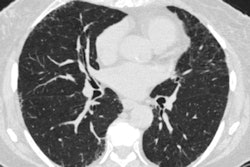
Volumetric CT imaging for lung cancer patients shows promise in this regard, so Xu and colleagues conducted a study that included 100 patients with lung cancer, all of whom underwent volumetric CT. They analyzed imaging data for the volume of the inspiratory phase (Vin), mean inspiratory lung density (MLDin), the volume of the expiratory phase (Vex), and mean lung density at the expiratory phase (MLDex). The team then used pulmonary function exam and volumetric CT analysis results to predict postoperative lung function.
Overall, Xu's group found that correlations between CT imaging data and pulmonary function exam results were significant, both pre- and postlung cancer surgery. It also found the following:
- Forced vital capacity, forced expiratory volume in the first second of imaging, and the ratio of these two estimated by volumetric CT analyses showed high concordance with those identified on pulmonary function examination.
- Preoperative volume of the inspiratory phase and volume of expiratory phase and mean lung density at expiratory phase and mean inspiratory lung density values proved to be predictors for postsurgery complications (hazard ratios, 5.39 and 6.52, respectively).
| Analysis of risk factors for post-lung cancer surgery complications | ||
|---|---|---|
| Parameter | Hazard ratio | p-value |
| Preoperative MLDex-MLDin | 6.52 | 0.008 |
| Preoperative Vin-Vex | 5.39 | 0.014 |
| Age | 3.56 | 0.079 |
| Clinical stage | 3.51 | 0.077 |
| Smoking history | 2.35 | 0.179 |
| Gender | 1.4 | 0.608 |
The study findings underscore not only radiology's part in managing lung cancer care but also CT imaging's, according to the authors.
"Radiology plays an extremely important role in the management of cancer patients, as it can accurately describe the condition and organ status of cancer in detail thus assisting in treatment plan making," they concluded. "The lung function assessment technology derived from CT technology will undoubtedly have its unique technical advantages and operability."
The complete study can be found here.